
Unmasking the Tiny Culprits: How New Tech Reveals Hidden Activity in Stroke Damage

"Laser-scanning nano-zymography offers a powerful new way to study microparticles and their role in stroke recovery, potentially revolutionizing diagnostic and treatment strategies."
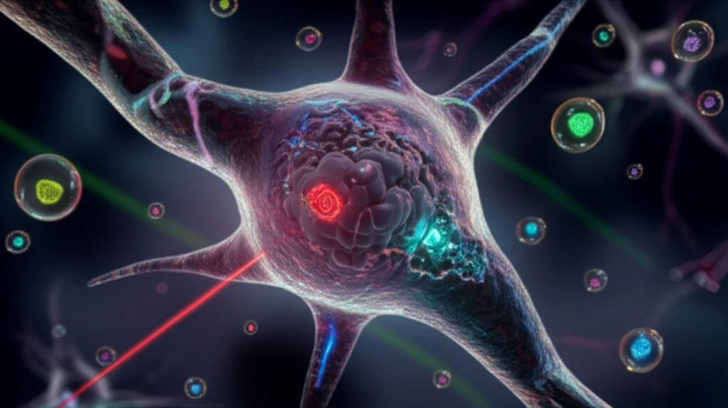
Imagine tiny bubbles, far smaller than the width of a human hair, carrying messages and tools between cells in your body. These are microparticles (MPs), and they're constantly released by cells to communicate and influence their environment. In the context of stroke, these MPs can either contribute to further damage or, potentially, aid in recovery. Understanding their role is crucial for developing more effective treatments.
The challenge has always been seeing what these MPs are actually doing. They're incredibly small, move rapidly, and are diverse in their composition. Traditional methods struggle to accurately measure their size, number, and, most importantly, their biological activity.
Now, researchers have developed a groundbreaking technique called nano-zymography, which utilizes laser-scanning confocal microscopy to directly image and functionally characterize these MPs. This method allows scientists to capture MPs, measure their size, detect specific surface markers, and even evaluate their proteolytic activity – their ability to break down proteins. This opens up exciting possibilities for understanding the complex role of MPs in stroke and other diseases.
The Hidden Toll of Stroke
Stroke remains one of the leading causes of death and long-term disability worldwide, with millions affected each year. While mortality rates have improved due to advances in acute care, many survivors continue to face significant neurological impairments. The economic burden on healthcare systems and patients alike underscores the urgent need for better diagnostic and therapeutic approaches. Understanding the full scope of stroke's impact requires ongoing research into both prevention and recovery strategies.
Current Treatment Challenges
Acute ischemic strokes represent a major health concern, with intravenous thrombolysis established as the main treatment for over three decades and endovascular therapy emerging as a subsequent advancement. Despite these interventions, most patients remain disabled following treatment. Nanomedicine has shown considerable promise in stroke management, yet imaging modalities face technical limitations in signal sensitivity and tissue penetration. Current nanomedicine approaches for thrombolytic therapy show potential for reducing infarct volume and improving neurological outcomes in preclinical studies.
The Evolution of Nanotechnology in Stroke Care
The term nanotechnology refers to science conducted at the nanometer scale, involving structures with dimensions between 1 and 100 nanometers. Over the past two decades, nanoneuroscience and nanoneurosurgery have emerged as new frontiers in biomedical science and clinical practice. Nano and micro-technologies are utilized for therapeutic delivery of biologics and small molecules in formulations ranging from one nanometer to 100 microns or more. These technologies possess unique physiochemical properties that have enabled astonishing advancements in stroke research and treatment development.
Nano-Zymography: A High-Resolution Look at Microparticle Activity
The new technique hinges on a clever method of trapping MPs. Researchers coat tiny microwells with annexin-V, a protein that binds strongly to phosphatidylserine (PS), a molecule found on the surface of most MPs. This essentially creates a sticky surface that captures and immobilizes the MPs, preventing them from drifting out of view.
- Measuring Size and Number: A fluorescent dye called CFSE labels virtually all cell-derived MPs, allowing for accurate counting and size measurements using specialized image analysis software.
- Detecting Surface Antigens: Specific antibodies, tagged with fluorescent markers, can be used to identify proteins on the MP surface, revealing their origin and potential function.
- Evaluating Proteolytic Activity: This is where the "zymography" part comes in. Researchers use fluorescent substrates that are cleaved by specific enzymes (proteases). If an MP has proteolytic activity, it will break down the substrate, releasing a fluorescent signal that can be detected and measured.
Cutting-Edge Nanomaterial Applications
Recent advances in nanomaterials present new opportunities in stroke detection, diagnosis, and treatment. Researchers are exploring the utilization of nanomaterials across computed tomography, magnetic resonance imaging, and other diagnostic modalities. These innovations aim to improve clinical diagnosis of stroke patients while elucidating the potential of nanotechnology in post-stroke therapy. Future research directions and potential clinical applications continue to be identified as this field rapidly evolves.
Challenges and Limitations in Nanomedicine
Despite the promise of nanotechnology in stroke treatment, significant challenges remain in translating preclinical successes to clinical applications. Variability in study designs and methodologies complicates the assessment of nanomedicine's true effectiveness. Safety concerns and potential side effects of nanoparticle-based therapies require thorough investigation before widespread clinical adoption. The gap between laboratory research and real-world implementation continues to pose substantial hurdles for this emerging field.
Stage-Based Nanotechnology Approaches
Ischemic stroke management can be categorized into four stages: prevention, neuroprotection, revascularization, and adjunctive therapy. Each stage involves distinct pathological mechanisms that require different nanotechnology-based therapeutic strategies. Nanotechnology enables spatiotemporal control across these stages, addressing the unique challenges presented at each phase of stroke progression. This stage-based framework helps researchers and clinicians tailor interventions to specific pathological conditions.
A Promising Tool for Stroke Research and Beyond
The development of nano-zymography represents a significant advancement in the study of microparticles and their role in disease. By providing a direct and detailed view of MP activity, this technique has the potential to unlock new insights into the complex mechanisms underlying stroke and other conditions.
Integrating Nanotechnology into Stroke Care
The integration of nanotechnology into stroke care represents a paradigm shift in how researchers approach both diagnosis and treatment. Expert consensus suggests that while significant progress has been made, substantial work remains to optimize nanoparticle-based interventions. Collaborative efforts between neuroscientists, engineers, and clinicians will be essential to overcome existing barriers. The field continues to evolve rapidly, with new discoveries regularly expanding the possibilities for patient care.
Nanozymes and Emerging Therapeutic Directions
Nanozymes have demonstrated outstanding achievements in recent years, particularly in the development of biosensors and therapeutic applications for ischemic stroke. These nanomaterial-based catalysts offer unique mechanisms for targeting stroke pathology at the molecular level. Research continues to illuminate the internal mechanisms and catalytic principles that make nanozymes effective therapeutic agents. The construction of nanozyme-based systems represents a promising frontier for next-generation stroke treatments.
Navigating the Path Forward
The broader healthcare system faces significant challenges in adopting nanotechnology-based stroke interventions, including regulatory hurdles and cost considerations. Infrastructure requirements for advanced nanomedicine manufacturing and administration present additional barriers to widespread implementation. Training healthcare professionals in these new technologies will be crucial for successful integration into clinical practice. Addressing these systemic challenges requires coordinated efforts across research institutions, healthcare providers, and policy makers.
From Laboratory to Patient Care
Ultimately, the success of nanotechnology in stroke care will be measured by its impact on patient outcomes and quality of life. Real-world implementation requires considerations beyond efficacy, including accessibility and affordability for diverse patient populations. Patient perspectives and experiences should inform the development and deployment of these advanced therapeutic approaches. The human element remains central to ensuring that technological advances translate into meaningful improvements in stroke recovery and rehabilitation.
The ability to identify and characterize fibrinolytic MPs in stroke patients is particularly exciting. These MPs, carrying tissue-type plasminogen activator (tPA), may play a crucial role in breaking down blood clots and restoring blood flow to the brain. Nano-zymography could help to identify patients who would benefit most from tPA therapy and to develop new strategies to enhance its effectiveness.
While further research is needed, nano-zymography holds great promise as a valuable tool for both experimental and clinical studies, potentially leading to more effective diagnostic and therapeutic interventions for stroke and a range of other diseases.