
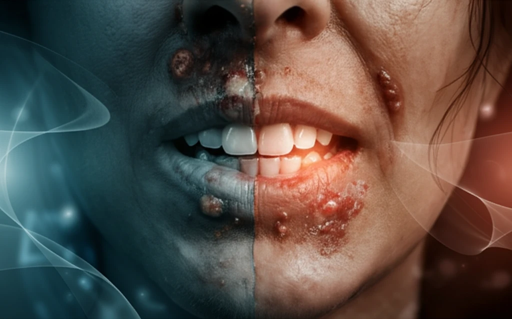
Unmasking Oral Mucosal Lesions: A Comprehensive Guide to Symptoms, Prevalence, and Expert Insights

"Navigating the complexities of oral health: Delve into our guide on oral mucosal lesions (OMLs), understanding their symptoms, prevalence, and latest research for better oral care."
Oral mucosal lesions (OMLs) are a widespread concern, impacting countless individuals worldwide. These lesions, which manifest as sores, ulcers, or abnormal tissue changes in the mouth, can range from minor annoyances to indicators of more serious underlying health conditions. Despite their prevalence, OMLs are often underestimated in terms of their impact on quality of life and overall well-being. Recognizing the symptoms and understanding the risk factors are crucial first steps in managing and preventing these conditions.
The mouth is a gateway to the body, and its condition can reflect systemic health issues. Factors such as poor oral hygiene, tobacco use, excessive alcohol consumption, and certain medical conditions can all contribute to the development of OMLs. Moreover, these lesions can cause significant discomfort, affecting eating, speaking, and even self-esteem. Therefore, a proactive approach to oral health is essential, involving regular check-ups with dental professionals and adopting healthy lifestyle habits.
This article aims to shed light on the multifaceted nature of OMLs. We will explore the prevalence of these lesions in various populations, dissect the common symptoms, and provide insights from expert research. By understanding the causes, treatments, and preventive measures, individuals can take control of their oral health and minimize the impact of OMLs on their daily lives. Let's embark on a journey to unravel the complexities of oral mucosal lesions, empowering you with the knowledge to maintain a healthy, confident smile.
Common Lesions and Their Clinical Impact
The most common superficial oral mucosal lesions include candidiasis, recurrent herpes labialis, recurrent aphthous stomatitis, erythema migrans, hairy tongue, and lichen planus, according to the American Academy of Family Physicians. Beyond these everyday presentations, research in inflammatory bowel disease patients found that oral mucosal lesions and oral symptoms were positively associated with disease activity, conceivably linked to corticosteroid and immunosuppressant therapy. Hospital-based studies have also assessed oral mucosal lesions alongside adverse habits using the World Health Organization's 1980 criteria, underscoring how behavioral factors are part of the standard evaluation. Epidemiological work further examines how oral mucosal lesions correlate with dermatological preliminary diagnoses, reflecting the broad, cross-specialty impact of these conditions.
Diagnosis, Dermoscopy, and the Limits of Symptom-Based Care
Traditional management of oral mucosal lesions often focused on symptomatic relief and lesion excision, with limited emphasis on the underlying etiological factors. Diagnosis is complicated by the fact that white lesions frequently arise from a thickening of the surface keratin layer through traumatic, inflammatory, or neoplastic pathways, which can present in similar ways. Although dermoscopy has been proposed as an aid, it is technically challenging for oral lesions because of the anatomical configuration of the oral cavity, the limited availability of dedicated mucosal probes, and difficulties achieving stable contact with moist surfaces. Because pharmacists are often among the first points of contact for people with oral pain and lesions, their initial management decisions can shape the course of care.
Epidemiological Foundations and Enduring Classifications
The study of the oral mucosa has a long history in medicine and dentistry, with early work establishing the basic architecture and classification of the oral lining. Modern epidemiological studies continue to refine this picture: in one Brazilian cohort, 465 lesions were diagnosed in 376 (46.9%) of participants, illustrating just how frequently oral mucosal lesions occur in examined populations. Notably, the presence of actinic cheilitis was significantly associated with fibrous inflammatory hyperplasia and low literacy in the same cohort. These findings align with the broader recognition that oral mucosal lesions are quite common, with some types more prevalent in certain groups than in others.
Understanding Oral Mucosal Lesions: Prevalence and Symptoms
Recent research conducted in Sweden provides valuable insights into the prevalence and characteristics of oral mucosal lesions (OMLs) in an adult population. The study, which involved over 6,000 participants, revealed that approximately 14.7% of individuals presented with some type of OML. This highlights the significant burden of these conditions on public health, underscoring the need for greater awareness and proactive management.
- Snuff dipper's lesion: Often appears as a white or grayish patch in the area where smokeless tobacco is placed.
- Lichenoid lesions: These lesions can manifest as white, lacy patterns or raised plaques on the oral mucosa, often accompanied by soreness or burning sensations.
- Geographic tongue: Characterized by irregular, map-like patterns on the surface of the tongue, which may shift over time.
New Population Studies and Emerging Risk Factors
Recent research has expanded the evidence base for oral mucosal lesions beyond clinic-based samples. A retrospective study of a Chilean elderly population found that oral mucosal lesions and normal variations of oral anatomy are very common in this group, yet few studies have examined their frequency worldwide, and fewer still in Chile. Similarly, a Norway-focused study noted that most prevalence research on oral mucosal lesions centers on patient populations, and it set out to estimate prevalence in a broader sample. A scoping review has also begun systematically mapping the oral cavity lesions reported in people who smoke electronic cigarettes, reflecting growing interest in e-cigarettes as an emerging factor in oral mucosal disease.
Unanswered Questions and Limitations in the Evidence
It is worth noting that the evidence base on oral mucosal lesions carries real limitations. Much of the published literature is drawn from specific clinical, hospital, or elderly populations, which may not reflect the general population, and the underlying causes of many lesions remain incompletely understood. Because lesions of very different origins can look alike, misdiagnosis remains a practical risk in everyday care. Readers should therefore treat prevalence and outcome figures with some caution until larger, more representative studies provide definitive data.
Age-Related Changes Versus Benign Mimics
Oral mucosal lesions are not a single disease but a spectrum, and age is a recognized contributing factor: halitosis (bad breath), xerostomia (dry mouth), and periodontal disease are oral conditions that can lead to mucosal lesions with advancing age. At the same time, oral lesions can closely resemble one another, which is why clinicians must be able to recognize their distinctive features. Distinguishing benign from malignant disease, and deciding when a biopsy is warranted, are central to that evaluation. Comparing these angles shows that while aging creates vulnerability, careful differential diagnosis remains the key safeguard against both overtreatment and missed malignancy.
Taking Control of Your Oral Health
Understanding oral mucosal lesions (OMLs) is essential for maintaining overall health and well-being. By recognizing the symptoms, seeking timely diagnosis, and adopting preventive strategies, individuals can minimize the impact of these conditions on their daily lives. Regular dental check-ups, good oral hygiene practices, and awareness of risk factors such as tobacco and alcohol use are all important steps in protecting your oral health. Armed with knowledge and proactive care, you can ensure a healthy, confident smile for years to come.
Expert Consensus on the Gold Standard
Within oral medicine, histopathological examination remains the gold standard for the diagnosis of oral mucosal lesions, according to an American Journal of Oral Medicine report. Despite this reliance on biopsy-based diagnosis, little is known about the level of clinical-pathologic agreement for lesions diagnosed by oral medicine experts. That gap matters because expert clinical impressions are used routinely in triage and in research settings. It underscores the value of combining careful clinical examination with histopathology rather than treating the two as interchangeable.
Building Surveillance for the Long Term
Trend data on oral mucosal lesions are limited, and the literature is composed largely of prevalence studies documenting lesions in different populations. Reviews of the available data on leukoplakia, recurrent herpes labialis, and recurrent aphthous ulcers have outlined strategies for monitoring these conditions and the factors that may affect future trends. Establishing standardized surveillance would allow researchers to detect shifts in lesion patterns over time. Both reviews point in the same direction: consistent, comparable data collection is the foundation for understanding where oral mucosal disease is headed.
The Mouth as a Window to the Body
Oral mucosal lesions are frequently the oral manifestations of systemic diseases, and recognizing them carries diagnostic significance that extends beyond the mouth itself. A chapter on systemic diseases with oral mucosal manifestations emphasizes both their diagnostic value and their clinical management. For clinicians, this means oral findings can serve as early or corroborating clues to an underlying condition. Managing such lesions therefore requires coordination between oral health providers and the physicians treating the systemic disease.
Quality of Life at the Center of Care
Oral mucosa lesions do not just present clinical challenges; they also negatively impact the quality of life of the patients who suffer from them. A cross-sectional study in Sudan found that oral mucosal lesions and oral symptoms influence oral health-related quality of life in dermatological patients. This human dimension matters because persistent pain, difficulty eating, or anxiety about a lesion can weigh heavily on daily life even when the lesion itself is benign. It is a reminder that successful treatment should be measured not only in tissue outcomes but in how patients feel and function.