
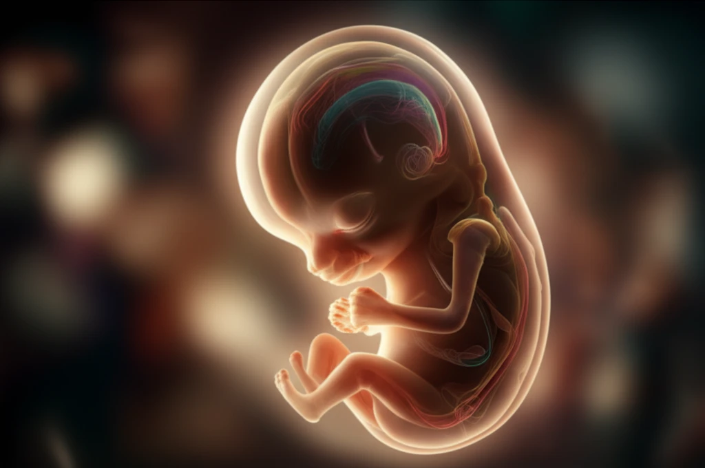
Unlocking the Secrets of Life: How Scientists Are Mapping the Body's Blueprint for Growth

"From Tiny Embryos to Complex Tissues: A Groundbreaking Study Reveals the Hidden Mechanics Behind How Our Bodies Take Shape."
Imagine the incredible journey from a single fertilized egg to a fully formed human being. It's a process of astonishing complexity, orchestrated by a symphony of biological processes. Scientists have long sought to understand the intricate dance of cell division, tissue formation, and overall growth that shapes us from the very beginning. A new study has delved deep into this microscopic world, providing a fresh perspective on how our bodies develop.
At the heart of this research lies the concept of morphogenesis – the biological process that causes an organism to develop its shape. This involves cell movements, tissue rearrangements, and changes in cell behavior that collectively sculpt the body's structures. This research looks at this in the early stages of development. By focusing on the early stages of development, the scientists have illuminated the fundamental mechanisms that drive this essential process.
This research, published in Development Advance Online Articles, provides a detailed analysis of the mechanics of embryonic axis elongation – the process by which the basic body plan is established. The research team used advanced imaging techniques and computational analysis to track cell behavior and tissue deformations in developing quail embryos. Their findings offer a window into the fundamental principles that guide the development of complex biological systems.
Why the Blueprint Matters
Mapping embryonic development helps scientists understand how a fertilized egg becomes an organized body. This knowledge may eventually clarify why development succeeds, why it sometimes fails, and how early biological decisions shape later health. Because research remains ongoing, its clinical impact should be described as promising rather than fully established.
Studying Development
Researchers study human embryonic development through embryo culture, transcriptional profiling, lineage mapping, and clinical observation. In IVF, embryologists commonly assess embryo morphology when selecting an embryo for transfer, but that judgment does not guarantee viability or sustained development in utero. Implantation remains a major black box because it occurs inside the uterus and is difficult to observe directly, while ethical and physical limitations restrict research. Recent methods have expanded access to early developmental processes, but important technical limitations remain.
From Preformation to Development
Embryology developed from early attempts to explain how embryos form and change over time. In the 17th century, some embryologists supported preformationism, the belief that embryos were miniature pre-existing adults; the theory was later challenged by William Harvey and others. Modern embryology instead describes development through processes including cell division and cellular differentiation. Educational timelines now organize human development week by week, linking approximate timing to major developmental events.
Unraveling the Mysteries of Embryonic Growth: Key Findings from the Groundbreaking Study
The research team focused on understanding how different tissues coordinate their growth during the elongation of the embryo's axis. This process is crucial for forming the posterior part of the body, including the spine and other vital structures. The scientists employed advanced imaging techniques, including two-photon laser imaging, to observe the behavior of cells within the developing quail embryos. They examined processes such as cell proliferation and apoptosis, which play vital roles in tissue growth and development.
- Differential Growth Rates: The study revealed that different tissues grow at distinct rates, contributing to the complex shaping of the embryo.
- Cell Density and Size: The research indicated that tissue expansion during elongation isn't solely due to changes in cell density or size, but primarily from the addition of new cells.
- Proliferation and Apoptosis: The study highlighted the importance of cell cycle times and cell death in determining the pace of growth of various tissues.
A Rapidly Advancing Field
Recent work is expanding the ability to study human embryonic development in greater detail. Researchers are developing improved culture methods, molecular profiling approaches, and tools for tracing cell lineages during early development. These advances may reveal mechanisms that were previously difficult to observe, although the field remains technically challenging and many findings are still being translated into broader biological or clinical understanding.
Why Development Fails
Human embryos often struggle to cope with high levels of replication stress early in development, a problem that can impair fertility treatment and may have longer-term health implications. Embryo development failure is also described as common in IVF and can result from multiple biological causes, rather than from anyone’s individual fault. Researchers studying early human development face technical obstacles that complicate both observation and interpretation. Developmental defects can involve structural or functional abnormalities arising from fertilization through early organ and tissue formation.
Human and Animal Models
Comparative embryology uses shared staging systems, including Carnegie stages, to examine similarities and differences among human and animal embryos. Workshop discussions emphasize that nonhuman embryos, embryo models such as chimeras, and human embryos can answer overlapping but not identical scientific questions. Important differences may involve fertilization location, embryonic layer formation, and placental development. Mouse research remains useful, but comparisons with humans must account for species-specific developmental differences.
Looking Ahead: Implications and Future Directions of the Research
This research provides a detailed look into the mechanics of embryonic development. The study's findings not only deepen our understanding of fundamental biological processes but also open doors for future research. By continuing to explore the cellular and molecular mechanisms behind tissue formation and axis elongation, scientists hope to gain a more comprehensive understanding of the human body's complex blueprint, which can pave the way for advances in regenerative medicine, and treating developmental disorders. The journey from a single cell to a complete organism continues to fascinate and inspire, and this research marks an important step forward in unraveling its secrets.
Models and Their Limits
Stem cell-based mammalian embryo models make developmental mechanisms easier to investigate because they are more amenable to genetic and epigenetic perturbations than natural embryos. Recent models have produced a wide range of experimental systems for studying embryonic development. Their imperfections are scientifically important because they expose gaps in current understanding rather than providing complete replicas of natural embryos. Public and expert opinions also vary on research involving induced-pluripotent-stem-cell embryo models and cultures beyond or within 14 days.
Opening the Black Box
Future embryonic-development research is expected to pursue a better understanding of human biology, with the potential to prevent or treat malformations and serious health conditions. A model described by Yale researchers offers a window into gastrulation, the stage two to three weeks after conception when embryonic cells begin becoming specialized and primitive body axes emerge. Embryoids may broaden experimental possibilities, but their developmental potential raises distinct ethical questions. Researchers therefore face both a scientific opportunity and a need to clarify how these models relate to sexual embryos.
Beyond the Embryo
Embryonic and fetal development must be understood within the broader physiological and clinical context of the mother. Gene editing in embryos or fetuses presents a potential rationale when disease begins early, permanent damage may occur during critical developmental periods, or immune responses could complicate treatment. However, human embryo genome editing still faces concerns about off-target effects, mosaicism, and profound ethical questions. Comprehensive safety protocols remain necessary before clinical implementation can be considered.
Science, Treatment, and Ethics
Embryology has contributed to fertility treatment and genetic research, giving developmental biology consequences beyond the laboratory. Embryo selection research can compare conventional light-microscopic examination with time-lapse imaging, but selecting and transferring an embryo raises significant ethical issues because it may affect a future person. Genome editing has also been proposed to correct inherited mutations, including a mutation associated with hypertrophic cardiomyopathy that can cause heart failure. These applications show why advances in embryo research require both medical evaluation and careful ethical scrutiny.