
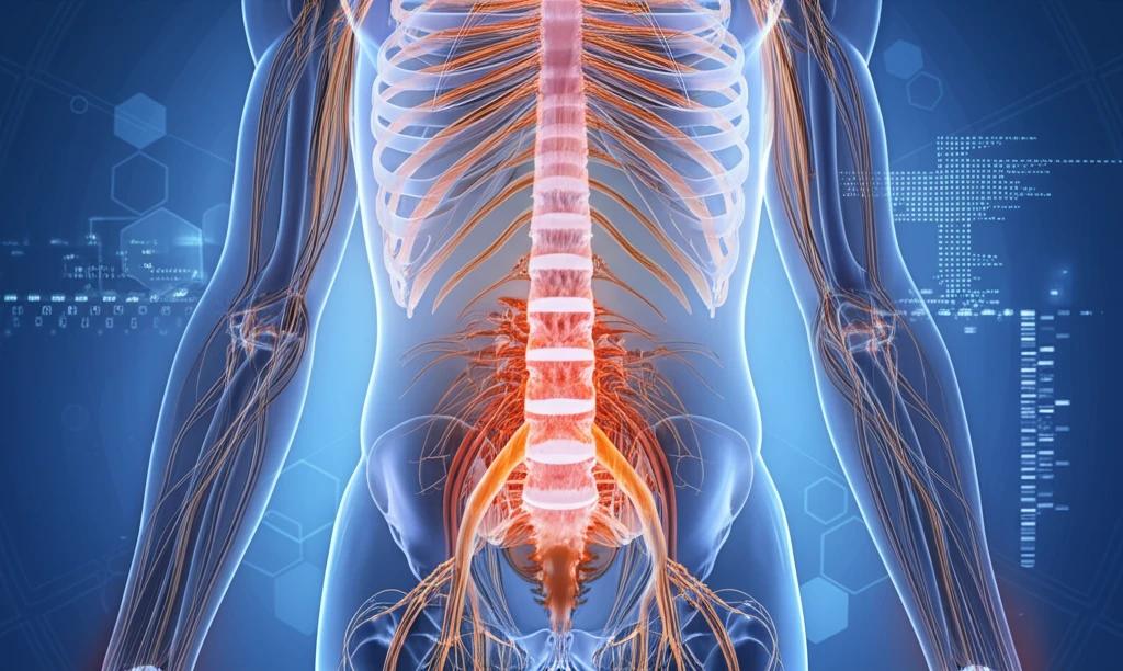
Digital illustration of a human spine with intricate nerves and tissue, representing osteolipomas.

Unlocking the Mystery of Lumbar Intraspinal Osteolipomas: A Comprehensive Guide

"Rare Spinal Tumor Explained: Cauda Equina Syndrome, Diagnosis, and Hope for Recovery"


Lipomas, benign mesenchymal tumors, are common, but osteolipomas are a rare variant, constituting only 1% of all lipomas. These unusual formations typically appear in the oral cavity, brain, and neck, with scarce occurrences in the knee and thigh. Intraspinal osteolipomas are exceptionally rare, with only a few documented cases, mainly in the cervical and thoracic spine. A new case of osteolipoma in the lumbar spine has recently been documented.

This comprehensive guide explores a recent case of a solitary lumbar intraspinal osteolipoma presenting as cauda equina syndrome. The tumor was successfully excised, and spinal fusion was performed to stabilize the spine. This detailed exploration covers the patient's clinical presentation, diagnostic journey, treatment course, and histopathological findings, combined with a thorough review of existing literature.

Osteolipomas, while benign, can cause rapid neurological decline, making prompt diagnosis and treatment critical. Understanding the nuances of these rare tumors can significantly improve patient outcomes. This article aims to shed light on lumbar intraspinal osteolipomas, their diagnosis, treatment, and the potential for recovery.

AI Search Multiple angles on this topic

Lumbar Anatomy and Clinical Relevance

The lumbar region, often called the lower spine, contains five vertebrae. These vertebrae are the largest and strongest movable bones in the spinal column, designated L1 through L5. The lumbar spine lies below the 12 thoracic vertebrae and above the five fused bones of the sacrum. It includes vertebral bones, discs, nerves, muscles, ligaments, and blood vessels, while the cauda equina nerve roots descend through the lumbar spinal canal after the spinal cord ends near its upper portion.

Anatomy-Guided Evaluation

A standard approach to lumbar conditions begins with a detailed understanding of the lumbar spine's anatomy. This includes the vertebral bodies, discs, nerves, and supporting muscles. Such anatomical knowledge is described as essential for identifying and addressing common disorders. However, the available source does not specify particular diagnostic methods or quantify their limitations, so conclusions about accepted procedures should be made cautiously.

An Evolving Field

The historical development of knowledge about lumbar intraspinal osteolipomas is not established by the supplied source material. In general, understanding of these lesions would be expected to evolve alongside advances in spinal anatomy, imaging, and surgical care. Specific milestones, dates, and foundational discoveries should therefore be verified against dedicated historical and clinical literature.

What are Osteolipomas and Why are They So Rare?

Digital illustration of a human spine with intricate nerves and tissue, representing osteolipomas.

Osteolipomas are unusual tumors composed of mature fat tissue combined with bone. Typically, lipomas are already common, but osteolipomas represent a tiny fraction of these, making them a rare and intriguing medical phenomenon. Most reported cases involve locations such as the oral cavities, head, brain, knee, and thigh. Spinal osteolipomas are exceptionally scarce, with lumbar occurrences being the least common.

The limited understanding of osteolipomas contributes to diagnostic challenges and treatment strategies. These tumors' rarity means that medical professionals may not immediately recognize them, potentially leading to delays in diagnosis and appropriate management. Early detection and accurate diagnosis are crucial to prevent neurological deterioration and ensure the best possible outcomes.

Here's a closer look at what makes osteolipomas unique:
  • Composition: A mix of mature fat and bone tissue.
  • Rarity: Only 1% of all lipomas are osteolipomas.
  • Location: Predominantly found in the oral cavity, head, brain, knee, and thigh.
  • Spinal Involvement: Extremely rare, especially in the lumbar region.
AI Search Multiple angles on this topic

Current Evidence

The supplied source material does not include recent studies or reviews focused specifically on lumbar intraspinal osteolipomas. A reliable assessment of current research would require evidence from contemporary case series, imaging studies, and systematic reviews. Without those sources, claims about research trends, outcomes, or emerging techniques would remain provisional.

Limits of the Evidence

The available material does not document specific counterarguments, failed approaches, or disputed findings concerning lumbar intraspinal osteolipomas. It consequently cannot support a detailed comparison of competing interpretations or treatment failures. Any such discussion should distinguish documented evidence from unresolved clinical uncertainty.

Comparative Perspective

No source supplied here compares lumbar intraspinal osteolipomas with other spinal lesions or treatment strategies. Meaningful comparative analysis would require matched information about anatomy, imaging characteristics, clinical effects, and outcomes. Those comparisons should not be inferred from the general lumbar anatomy information alone.

Several theories attempt to explain the development of osteolipomas, including repeated microtrauma, metaplasia of fibroblasts to osteoblasts, and chondroossification of preexisting mesenchymal cells. Genetic factors, such as translocations involving the 12q13-15 chromosome, have also been observed in some cases. Continued research is essential to fully understand the underlying causes and mechanisms driving the formation of these unusual tumors.

Hope and Future Directions

The successful management of lumbar intraspinal osteolipomas relies on early diagnosis, precise surgical techniques, and comprehensive postoperative care. While these tumors are rare, understanding their characteristics and potential impact is crucial for healthcare professionals. Further research into the causes and optimal treatment strategies will undoubtedly improve outcomes for patients affected by this rare condition.

AI Search Multiple angles on this topic

Evidence-Based Interpretation

The supplied material supports viewing the lumbar spine as a structurally complex region containing five large vertebrae and closely related neural and supporting tissues. It does not provide lesion-specific expert commentary on lumbar intraspinal osteolipomas. A responsible synthesis should therefore separate established lumbar anatomy from conclusions that require specialized clinical evidence.

Unresolved Directions

Future progress in understanding lumbar intraspinal osteolipomas will likely depend on more focused clinical and research evidence. Potential priorities include clearer characterization, improved diagnostic evaluation, and better documentation of outcomes, although the supplied sources do not establish specific future technologies or milestones. These possibilities should be treated as areas for investigation rather than settled predictions.

Context and Uncertainty

Lumbar disorders must be considered within a region that combines vertebrae, discs, nerves, muscles, ligaments, and blood vessels. This anatomical complexity can make lesion-specific interpretation clinically demanding. The supplied sources do not describe broader healthcare-system barriers, access problems, or population-level effects related specifically to osteolipomas.

Patient-Centered Importance

Conditions affecting the lumbar region can matter greatly to patients because the area supports the upper body and participates in movement. The lumbar canal also contains descending cauda equina nerve roots after the spinal cord ends. However, the supplied sources do not provide patient-reported outcomes or lesion-specific accounts of daily-life impact, so those effects should not be generalized beyond the available anatomical context.

Reading the findings from this case

The report links a compressed cauda equina and urinary and neurological symptoms with a surgically removed lesion. Improvement was reported over follow-up, but the motor findings were described as partial recovery, not a return to normal function.[1] [2]

This single case documents one patient's presentation and course. It cannot establish how often the same symptoms or recovery pattern occur in other people with this lesion.[2]

Limits of the lumbar case evidence

  • Plain spinal radiographs did not clarify whether the spine was unstable, leaving that assessment inconclusive in this case.[2]
Study at a glance
SettingOutpatient department[1]
DurationFollowed up for 2 years since surgical intervention[1]
Effect sizeAt 12 weeks, bilateral L4 and L5 motor power was MRC grade 3/5 and S1 root power was MRC grade 4/5[1]
Conflicts of interestNo conflicts of interest declared[1]
Times cited6[1]

How later spinal reports place this case

The 2018 paper described a lumbar intraspinal case as unprecedented in its literature review; later reports show that lumbar examples were subsequently documented, including an L3/4 lesion reported in 2025.[1] [3] [4]

A 2025 review counted four prior reports of intraspinal osteolipomas with osseous components, while a 2026 cervical case report identified ten cases near the spine, including cases in multiple spinal regions. These are different literature counts with different stated scopes, not a direct measure of incidence.[4] [5]

What the reports leave unresolved

  • Do the proposed mechanisms—microtrauma, cellular metaplasia, or chondroossification—explain how these lesions form, or are they only hypotheses?[1]
  • How consistently do MRI signal patterns and CT findings distinguish osteolipomas from other lesions across reported cases?[1] [4]
  • How do surgical extent and the risk of complications relate to neurological outcomes across lumbar cases?[1] [6]

Questions about diagnosis and symptoms

What did the bladder test show in the reported patient?

A postvoid bladder ultrasound measured 250 ml of residual urine, which the report interpreted as a neurogenic bladder.[2]

What symptoms brought the patient to care?

The patient reported a year of low back pain with claudication symptoms, one-sided numbness, and foot paresthesia.[2]

About this Article -

Written with AI assistance from published research, and reviewed by the Mystum team. See our About page for more information.

This article is based on research published under:

DOI-LINK: 10.1155/2018/1945149, Alternate LINK

Title: Lumbar Intraspinal Osteolipoma Presenting As Cauda Equina Syndrome: A Case Report And Review Of Literature

Subject: General Earth and Planetary Sciences

Journal: Case Reports in Orthopedics

Publisher: Hindawi Limited

Authors: S. Dilip Chand Raja, Rishi Mugesh Kanna, Ajoy Prasad Shetty, S. Rajasekaran

Published: 2018-10-22

Everything You Need To Know

1

What exactly are osteolipomas, and what makes them so uncommon compared to other types of tumors?

Osteolipomas are very rare, benign tumors composed of mature fat tissue and bone. While lipomas themselves are relatively common, osteolipomas only make up about 1% of all lipomas. They are most often found in areas like the oral cavity, head, brain, knee, and thigh. Their occurrence in the spine, particularly the lumbar region, is extremely rare. The limited number of cases documented contributes to the challenges in diagnosing and treating them effectively.

2

How can a lumbar intraspinal osteolipoma lead to Cauda Equina Syndrome, and why is this condition so critical to address?

Lumbar intraspinal osteolipomas can lead to Cauda Equina Syndrome due to the tumor compressing the nerves in the lumbar spine. Because osteolipomas, though benign, can cause rapid neurological decline, this compression can result in symptoms like lower back pain, bowel and bladder dysfunction, and leg weakness. This syndrome is an indication for prompt diagnosis and treatment of the osteolipoma.

3

What are the suspected causes behind the development of osteolipomas, and what role do genetic factors play?

Several theories attempt to explain the development of osteolipomas. These include repeated microtrauma, metaplasia of fibroblasts to osteoblasts, and chondroossification of preexisting mesenchymal cells. Genetic factors, like translocations involving the 12q13-15 chromosome, are also being investigated. The exact cause remains unclear, highlighting the need for further research to fully understand how these tumors develop.

4

What are the main components of managing lumbar intraspinal osteolipomas, and what future research directions are being explored?

The keys to successful management of lumbar intraspinal osteolipomas include early diagnosis, precise surgical techniques to remove the tumor, and comprehensive postoperative care. Spinal fusion may be performed to stabilize the spine after tumor removal. Further research into optimal treatment strategies is essential to improve outcomes for individuals affected by this rare condition.

5

What are the potential long-term consequences of leaving a lumbar intraspinal osteolipoma untreated, and how can these risks be mitigated?

If left untreated, the compression from a lumbar intraspinal osteolipoma can lead to permanent neurological deficits, such as chronic pain, impaired bowel and bladder control, and muscle weakness in the legs. These long-term consequences can significantly impact a person's quality of life. Early diagnosis and treatment are crucial to prevent these complications and improve the chances of a full recovery. Spinal instability, while not directly caused by the osteolipoma itself, can arise after surgical removal of the tumor, potentially requiring spinal fusion to stabilize the spine and prevent further neurological issues.

Newsletter Subscribe

Subscribe to get the latest articles and insights directly in your inbox.