
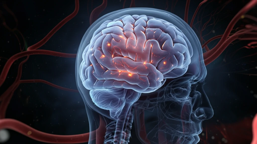
Unlocking the Enigma: High-Grade Angiocentric Glioma and the Quest for Early Diagnosis

"A Deep Dive into a Rare Brain Tumor and the Latest Imaging Techniques That Could Save Lives"
In the ever-evolving landscape of medical science, where breakthroughs often hinge on the ability to detect and understand anomalies, a spotlight shines on the intricate world of brain tumors. Among these, angiocentric glioma presents a particularly compelling challenge. Once considered a relatively benign entity primarily affecting young children, recent findings suggest a more complex reality, including the potential for aggressive, high-grade variants in older patients.
Angiocentric gliomas, now recognized as distinct neuroepithelial tumors by the World Health Organization (WHO), are characterized by their unique growth pattern around blood vessels. While traditionally associated with slow-growing, low-grade neoplasms causing chronic seizures in pediatric patients, the identification of high-grade forms necessitates a re-evaluation of diagnostic and treatment strategies. This article explores the subtle nuances of this rare tumor, highlighting the latest research and imaging techniques that promise earlier detection and improved patient care.
Imagine a scenario where subtle changes on an MRI scan can differentiate between a benign lesion and a potentially life-threatening malignancy. This is the frontier of modern neuroradiology, where advanced imaging modalities like diffusion imaging, spectroscopy, and tractography are transforming our understanding of complex brain tumors.
A Rare Tumor of Children and Young Adults
Angiocentric glioma is a rare type of brain tumor that primarily affects children and young adults and is classified as a low-grade glioma, generally slow-growing and less aggressive than other brain tumors. It is a rare, epilepsy-associated, low-grade neoplasm of the central nervous system that typically presents with medication-refractory seizures in children and young adults.
Diagnosis Through Neurosurgery and Advanced Imaging
Diagnosing angiocentric glioma involves a combination of neurosurgical procedures and imaging studies. In one reported case, a two-year-old patient with a histologically-confirmed WHO grade I angiocentric glioma was evaluated using both MRI and MR spectroscopy, illustrating the role of advanced imaging in confirming the diagnosis.
A Newly Codified Clinicopathological Entity
Angiocentric glioma was recently recognized as a distinct clinicopathological entity in the 2007 World Health Organization Classification of Tumours of the Central Nervous System. Prior to this codification, it was not formally classified as its own tumor type within the central nervous system.
Decoding Angiocentric Glioma: A Case Study
In a recent case that underscores the diagnostic challenges posed by angiocentric glioma, a 15-year-old male presented with progressive weakness and numbness on the left side of his body. Initial neurological examination revealed a range of symptoms, including bilateral papilledema, facial droop, and reduced strength in the extremities. Routine laboratory investigations yielded normal results, leading to an initial diagnostic dilemma.
- Advanced imaging techniques, including diffusion imaging, spectroscopy, and tractography, are transforming our understanding of complex brain tumors.
- MR spectroscopy revealed elevated choline/phosphocreatine and choline/N-acetyl aspartate ratios, suggesting an aggressive tumor.
- The case underscores the diagnostic challenges posed by angiocentric glioma.
- DTT aided in neurosurgical planning by mapping neuronal fiber tracts.
First Illustrated MR Spectroscopic Profile
Researchers have presented MRI and MR spectroscopy features from a two-year-old patient with a histologically-confirmed WHO grade I angiocentric glioma, noting that the spectroscopy profile of a typical case had not been previously illustrated. Another clinicopathologic review of five tumors described angiocentric glioma as a rare, epilepsy-associated, low-grade neoplasm with a characteristic perivascular growth pattern.
Diagnostic Challenges and Mimicry
Angiocentric glioma can mimic other conditions, as illustrated by a reported case in which the tumor presented like encephalomalacia on imaging, potentially complicating and delaying diagnosis. Brain tumor symptoms also differ depending on the area of the brain affected, ranging from very general complaints such as headache to very specific changes such as vision changes, which makes early recognition challenging.
Low-Grade Nature Compared With Other Brain Tumors
Angiocentric glioma is classified as a low-grade glioma, meaning it is generally slow-growing and less aggressive compared with other brain tumors. While brain tumors are rare relative to all cancers worldwide, the location of the tumor determines how it is treated, and symptoms vary based on the specific region of the brain affected.
The Future of Angiocentric Glioma Diagnosis
This case serves as a potent reminder that angiocentric glioma, even in its high-grade form, should remain on the radar of clinicians evaluating brain tumors, particularly in adolescent and young adult patients. By integrating advanced imaging techniques with careful histopathological analysis, the medical community can strive toward earlier, more accurate diagnoses, ultimately improving outcomes for those affected by this rare and challenging neoplasm.
A Distinct Entity With Unique Features
Angiocentric glioma is a rare brain tumor mainly occurring in children and adolescents, with unique cellular and molecular characteristics. It is recognized as a distinct clinicopathological entity with a characteristic perivascular growth pattern and a strong association with epilepsy.
Toward Earlier Recognition and Better Data
Key areas of angiocentric glioma advocacy include raising public awareness about the condition to facilitate early diagnosis. Integrated disease databases that combine associated genes, mutations, phenotypes, pathways, and drugs from multiple data sources may help accelerate research and understanding of this rare tumor.
Awareness and Diagnosis as Systemic Hurdles
Because symptoms can be very general, such as headache, or very specific, such as changes in vision, tumors are not always recognized early. Diagnosing angiocentric glioma requires a combination of neurosurgical procedures, and public awareness efforts are seen as central to facilitating earlier diagnosis and better outcomes.
Seizures That Resist Medication
Angiocentric glioma typically presents with medication-refractory seizures in children and young adults, meaning standard seizure treatments often fail to control symptoms. Because the tumor primarily affects younger patients and disrupts everyday life through persistent epilepsy, early diagnosis is critical to guiding timely treatment and improving quality of life.