
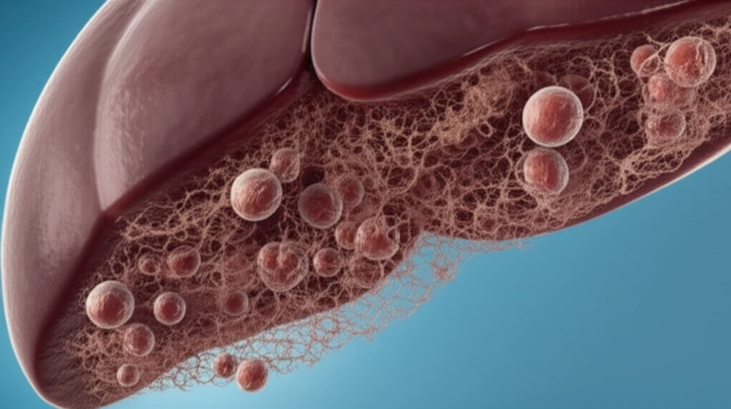
Unlocking Liver Fibrosis: How Parasites Offer Clues to Treatment

"Can understanding a rat parasite reveal the secrets to stopping liver disease in humans?"
Liver fibrosis, the scarring of the liver, is a serious health issue that can lead to cirrhosis and liver failure. Researchers are constantly seeking ways to understand how fibrosis develops and, more importantly, how to stop it. One intriguing avenue of investigation involves studying a specific type of liver fibrosis that occurs in rats infected with the nematode Capillaria hepatica.
In this model, rats develop septal fibrosis, a particular pattern of scarring in the liver, following infection with this parasite. What makes this model valuable is that researchers have observed that curative treatment of the infection can prevent the development of fibrosis if intervention occurs early enough – specifically, up to 15 days post-infection. However, treatment after this point is no longer effective in preventing fibrosis.
A recent study delved deeper into this phenomenon, aiming to pinpoint the parasitic factors present at the critical 15-day mark that determine whether or not septal fibrosis can be prevented. By understanding what's happening at this stage, scientists hope to uncover new targets for preventing and treating liver fibrosis in humans.
The Scale of Liver Disease
The CDC reports 4.5 million U.S. adults had diagnosed liver disease, equal to 1.8% of adults; those figures come from the 2018 National Health Interview Survey. Separately, an estimate cited for 2023 put new U.S. liver and intrahepatic bile duct cancer cases at 41,260, while 2022 saw 22,921 liver transplants in the United States. A 2025 systematic review and meta-analysis was conducted to evaluate the global prevalence and risk factors of advanced liver fibrosis and cirrhosis, but its provided summary gives no prevalence estimate.
Staging Fibrosis and Its Tradeoffs
Liver biopsy is commonly used to stage fibrosis and remains a reference standard for identifying and staging it. But biopsy is invasive, can involve sampling error and observer variability, and may need to be repeated. Performing biopsies at large scale may also be impractical or not cost-effective. These limitations have driven the development of noninvasive tests, some of which have better diagnostic value than biopsy in chronic liver diseases and are described as safe to perform.
The 15-Day Turning Point: What Happens When Treatment Fails?
The study focused on identifying which parasitic elements were present in the rats' livers at the point when curative treatment could no longer halt the progression of fibrosis. Researchers infected rats with Capillaria hepatica eggs and then treated different groups of rats with antihelminthic drugs (ivermectin and mebendazole) at various time points: 10, 12, 15, 17, and 20 days post-infection (PID).
- Both adult worms and eggs were present in the liver tissue.
- A complex necroinflammatory reaction (tissue death and inflammation) was occurring around the parasites.
- Importantly, no septal fibrosis was yet apparent.
Parasite-Related Fibrosis and Current Reviews
A report on granulomatous hepatitis caused by Calodium hepaticum in a captive mandrill in Brazil describes liver changes including parenchymal destruction, eosinophilic granulomas, and fibrosis. It notes that diagnosis remains challenging because of the parasite’s biological characteristics. Broader reviews of liver fibrosis cover its etiologies, diagnostic approaches, pathogenic mechanisms, antifibrotic agents in development, and future therapeutic directions. A Nature Reviews overview also highlights molecular and cellular mechanisms of hepatic fibrogenesis and their therapeutic implications.
A Narrow Window for Preventing Fibrosis
In rats infected with Capillaria hepatica, curative treatment prevented septal fibrosis when given through postinfection day 15, but not when begun later. This finding points to a timing limit in that experimental infection model; it does not establish the same treatment window in people. More broadly, liver fibrosis involves excessive extracellular matrix deposition and hepatic stellate cell activation, and may progress to cirrhosis and liver failure if left untreated. Chronic liver diseases with different causes can share these pathological features, underscoring that parasite-related findings do not by themselves resolve the wider challenges of fibrosis.
Eggs Hold the Key: Implications for Future Research
This research highlights the critical role of Capillaria hepatica eggs in driving the development of septal fibrosis in rats. While the initial damage may be triggered by the presence of adult worms and the associated inflammatory response, the continued presence of eggs seems to be essential for the fibrosis to progress.
Therapeutic Directions Ahead
A 2025 review describes liver fibrosis as a progressive consequence of chronic liver injury that can develop into cirrhosis, liver failure, or hepatocellular carcinoma. It identifies hepatic stellate cell activation, excessive extracellular matrix deposition, and structural disruption of liver tissue as features of fibrogenesis. The review discusses transforming growth factor-β among the processes involved, while the supplied summary offers no specific treatment results or projections. It frames ongoing treatment perspectives against the substantial global health burden of liver fibrosis.
A Difficult-to-Recognize Zoonotic Infection
The CDC identifies Capillaria hepatica, also called Calodium hepaticum, as the roundworm that causes hepatic capillariasis in humans; naming varies across regions and disciplines. The parasite is zoonotic, has low host specificity, and primarily occurs in rodent and carnivore hosts. Hepatic capillariasis can produce localized liver necrosis, fibrosis, and inflammation. A 2026 review describes the parasite as overlooked and says nonspecific clinical manifestations and diagnostic difficulty may lead to underestimation of its actual incidence.
A Child’s Diagnosed Infection
A case report described Capillaria hepatica infection in a four-year-old boy who had fever, hepatomegaly, peripheral eosinophilia, and noticeable weight loss. The diagnosis was made by finding a degenerated nematode in liver tissue on histological examination. Separately, an animal case report attributed an otter’s death to C. hepatica-associated hepatitis and capsular adhesions, and reported parasite-associated liver lesions in all cases reviewed. These reports illustrate documented illness in individual cases, rather than establishing how common such outcomes are.
These findings align with previous studies that showed that the eggs of C. hepatica play a key role in the pathogenesis of septal fibrosis of the liver, but the initiation process seems more complex and initiates only when the infection reaches full maturity.
This understanding could pave the way for new therapeutic strategies targeting the eggs themselves or the mechanisms by which they trigger fibrosis. Future research could focus on identifying specific molecules released by the eggs that stimulate the fibrotic process, potentially leading to the development of drugs that block these signals and prevent liver scarring. While the research was done on rats, further research could indicate that these findings are applicable in humans.