Surreal illustration of a fetal brain with disrupted cytoskeleton, symbolizing the impact of hypothyroidism on neurological development.

Thyroid Health and Development: How Hypothyroidism Impacts Your Baby's Future

"Uncover the critical link between thyroid function during pregnancy and your child's neurological development, and what you can do to ensure a healthy start."


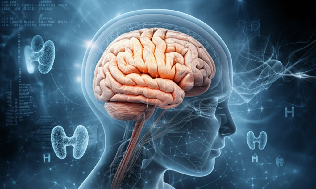
During pregnancy, thyroid hormones (THs) play a vital role in fetal development, particularly in the construction of the cytoskeleton – the structural framework within cells. This system, composed of microtubules (Tubulin), microfilaments (Actin), and intermediate filaments, is essential for neural cell shape, neuronal migration, and overall brain development. When thyroid hormone levels are insufficient, especially due to maternal hypothyroidism, the consequences can be far-reaching.

Research indicates that THs regulate and reorganize the cytoskeleton through non-genomic actions. These hormones also control the expression of the extracellular matrix (ECM) and adhesion molecules, crucial for neuronal migration and development. These include vital substances like tenascin-C, neural cell adhesion molecule (N-CAM), reelin and dab1, laminin, and fibronectin. These ECM and adhesion molecules are building blocks and must be available.

A mother's thyroid hormones directly influence the expression of genes related to neuronal migration, neurite branching, astrocytic cytoskeletal proteins, cell cycle regulators, neurotrophins and their receptors, and extracellular matrix proteins within the fetal brain. Ensuring adequate thyroid hormone levels is thus paramount for healthy neurological development.

The Chilling Effects of Hypothyroidism on Brain Development

Surreal illustration of a fetal brain with disrupted cytoskeleton, symbolizing the impact of hypothyroidism on neurological development.

Maternal hypothyroidism can significantly reduce the expression of Glial Fibrillary Acidic Protein (GFAP) in the fetal brain during late gestation. Studies on neonatal hypothyroid rats reveal disorganized actin fibers in the cerebellum, alongside disruptions in laminin. These abnormalities can alter cellular cytoskeleton development, impacting microtubule protein synthesis, axonal transport, neuronal outgrowth, and behavior. This disruption of axonal transport has downstream impacts.

These issues often stem from abnormalities in the cellular cytoskeleton, including the stabilization and composition of microtubule proteins and the delivery of these proteins to developing terminals via the slow component of axonal transport. This complex process is critical for proper brain formation and function.

  • Disrupted Cellular Structure: Hypothyroidism leads to disorganized actin fibers and disruptions in laminin, critical components of the brain's cellular structure.
  • Impaired Protein Synthesis: The condition affects the synthesis of microtubule proteins, vital for axonal transport and neuronal development.
  • Behavioral Changes: Resulting abnormalities impact neuronal outgrowth and behavior, leading to potential developmental delays.
Furthermore, early postnatal hypothyroidism can elevate both RNA and protein levels of tenascin-C in cerebellar Bergmann glia. Conversely, maternal hyperthyroidism can alter the expression of neuronal cytoskeletal proteins, accelerating fetal neuronal differentiation. Hypothyroidism reduces matrix glia protein (MGP) mRNA levels, while hyperthyroidism upregulates the MGP gene. Neurotrophins, like brain-derived neurotrophic factor (BDNF), crucial for development, are diminished in hypothyroidism and increased in hyperthyroidism. The activity of deiodinase 2 (D2) is also inhibited by T4 at the post-translational level, affecting microfilaments (Actin).

Future Directions: Can We Reverse the Damage?

The window for thyroid hormone-dependent regulation of these processes is primarily limited to pre- and perinatal life. Thyroid disorders during development can disrupt the cytoskeleton system, mitochondrial functions, and gene expression. Further research is needed to determine if normalizing circulating TH levels can prevent disturbances in fetal brain cytoskeletal protein expression. This could pave the way for interventions that mitigate the neurological impacts of thyroid imbalances during pregnancy, ensuring healthier outcomes for future generations.

About this Article -

This article was crafted using a human-AI hybrid and collaborative approach. AI assisted our team with initial drafting, research insights, identifying key questions, and image generation. Our human editors guided topic selection, defined the angle, structured the content, ensured factual accuracy and relevance, refined the tone, and conducted thorough editing to deliver helpful, high-quality information.See our About page for more information.

This article is based on research published under:

DOI-LINK: 10.4172/2161-1017.1000271, Alternate LINK

Title: Perinatal Hypothyroidism And Cytoskeleton Dysfunction

Subject: General Medicine

Journal: Endocrinology & Metabolic Syndrome

Publisher: OMICS Publishing Group

Authors: Ahmed Rg

Published: 2017-01-01

Everything You Need To Know

1

Why are thyroid hormones so important for a baby's brain development during pregnancy?

Thyroid hormones are vital during pregnancy because they directly influence the expression of genes related to neuronal migration, neurite branching, astrocytic cytoskeletal proteins, cell cycle regulators, neurotrophins and their receptors, and extracellular matrix proteins within the developing fetal brain. Adequate levels of thyroid hormones are essential for healthy neurological development.

2

What is the cytoskeleton and which components are critical for brain development influenced by thyroid hormones?

The cytoskeleton, which includes microtubules (Tubulin), microfilaments (Actin), and intermediate filaments, is crucial for neural cell shape, neuronal migration, and overall brain development. Thyroid hormones regulate and reorganize the cytoskeleton, and also control the expression of the extracellular matrix (ECM) and adhesion molecules like tenascin-C, neural cell adhesion molecule (N-CAM), reelin and dab1, laminin, and fibronectin, which are essential for neuronal migration and development.

3

How does maternal hypothyroidism impact the physical structure of the developing brain, and what are the potential consequences?

Maternal hypothyroidism can reduce the expression of Glial Fibrillary Acidic Protein (GFAP) in the fetal brain, leading to disorganized actin fibers in the cerebellum and disruptions in laminin. These abnormalities impact cellular cytoskeleton development, microtubule protein synthesis, axonal transport, neuronal outgrowth, and can even lead to behavioral changes. Proper axonal transport is crucial for delivering proteins necessary for brain formation and function.

4

Besides hypothyroidism, how does hyperthyroidism affect neuronal development, and what role do proteins like tenascin-C and neurotrophins play?

Early postnatal hypothyroidism can elevate the levels of tenascin-C in cerebellar Bergmann glia, while maternal hyperthyroidism can alter the expression of neuronal cytoskeletal proteins, potentially accelerating fetal neuronal differentiation. Additionally, hypothyroidism reduces matrix glia protein (MGP) mRNA levels, and hyperthyroidism upregulates the MGP gene. Neurotrophins, such as brain-derived neurotrophic factor (BDNF), are diminished in hypothyroidism and increased in hyperthyroidism, highlighting the delicate balance required for proper neurological development.

5

What future research is being conducted to potentially reverse or mitigate the negative impacts of thyroid imbalances on a developing baby's brain?

Research is focused on determining if normalizing circulating thyroid hormone levels can prevent disturbances in fetal brain cytoskeletal protein expression. This research aims to find interventions that could mitigate the neurological impacts of thyroid imbalances during pregnancy. Further investigation into the roles of factors like Glial Fibrillary Acidic Protein (GFAP), tenascin-C, matrix glia protein (MGP), and brain-derived neurotrophic factor (BDNF) is important to fully understand the potential for reversing or mitigating the effects of thyroid imbalances during pregnancy.

Newsletter Subscribe

Subscribe to get the latest articles and insights directly in your inbox.