
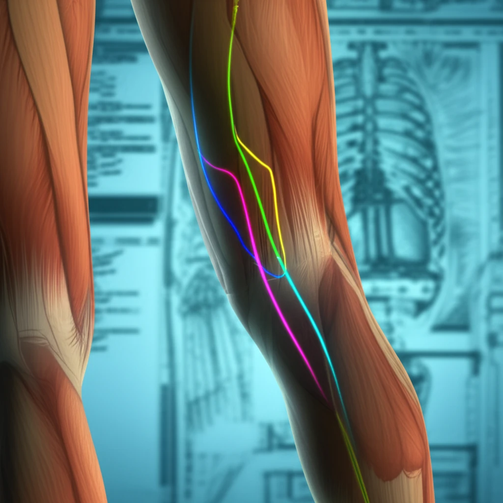
Sural Flap Anatomy: A Guide to Reliable Reconstruction

"Understanding the anatomical variations of the distally based sural flap for improved surgical outcomes."
Reconstructing soft tissue defects in the distal lower leg and foot presents a significant challenge due to limited local tissue availability and the complexities of the region. Free flaps, while an option, can be complex and carry potential drawbacks.
The distally based sural flap has emerged as a workhorse in reconstructive surgery for this area, offering a reliable solution for covering deep tissue defects. This fasciocutaneous flap relies on the sural artery and its perforators, making a thorough understanding of its anatomy crucial for successful outcomes.
This article delves into the anatomical intricacies of the sural flap, drawing upon cadaveric studies to provide surgeons with a detailed understanding of the flap's vascular supply and potential variations. By exploring these anatomical nuances, we aim to equip surgeons with the knowledge to optimize flap design and minimize complications.
Growing Use of the Reverse Sural Artery Flap
The reverse sural artery flap (RSAF) has emerged as a versatile option for soft tissue reconstruction in the distal lower extremity, particularly when microsurgical expertise or resources are limited. Despite its increasing use, comprehensive multicenter data on survival outcomes and anatomical site-specific performance remain limited. The RSAF is widely used for soft-tissue reconstruction of distal lower extremity defects, though outcomes may vary based on the defect site.
The Sural Flap as a Standard Reconstructive Option
Coverage of posttraumatic and chronic wounds at the distal leg is a difficult problem due to limited soft tissue available for local flaps. The sural flap remains a versatile and effective method for reconstruction in this area since it does not require microsurgical expertise. Perforator-based local flaps, such as propeller or keystone flaps, have made reconstruction more efficient while further reducing donor-site morbidity. The reverse sural artery flap is considered a standard method for reconstruction of soft tissue defects in the distal lower extremity.
Historical Development of the Sural Flap
The sural flap has its roots in early fasciocutaneous flap principles developed in the latter half of the twentieth century. Over time, surgeons refined the technique from pedicled designs to the reverse-flow configuration now commonly used. These foundational discoveries in vascular anatomy and flap hemodynamics laid the groundwork for modern reconstructive applications. The evolution of the procedure reflects broader advances in understanding of lower extremity perforasomes and angiosomes.
Unveiling the Sural Flap's Blood Supply: Key Anatomical Insights
A cadaveric study was conducted on eight lower limbs to meticulously map the arterial supply of the distally based sural flap. Red latex was injected into the popliteal artery, followed by careful dissection to reveal the fasciomyocutaneous flap and its vascular pedicle. The focus was on identifying and measuring the perforating arteries originating from the sural artery.
- Sural Nerve as a Guide: The sural nerve was consistently identified in all dissections, serving as a reliable landmark for locating the vascular pedicle.
- Perineural Plexus Dominance: In a significant number of cases, the primary blood supply to the flap originated from a perineural plexus surrounding the sural nerve.
- Arterial Variations: The study observed variations in the sural artery's presentation, including the presence of three distinct sural arteries (lateral, median, and medial) in some dissections, while others exhibited only lateral or combined lateral and medial arteries.
- Perforator Distribution: The number of perforating arteries varied, with 3 to 6 perforators identified on the medial side of the pedicle and 4 to 5 on the lateral side.
Contemporary Evidence on Sural Flap Outcomes
Recent studies have focused on evaluating long-term efficacy and surgical outcomes of sural neurovascular flap applications in distal lower limb wound reconstruction. Institutional comparative studies and systematic analyses of scientific evidence have been conducted to evaluate success and complication rates of classical and modified reverse sural flaps. There remains a need to investigate the impact of this reconstruction technique on patients' quality of life and esthetic satisfaction.
Risk Factors and Complications of Sural Flaps
Few studies have systematically assessed risk factors associated with poor outcomes in the distally based sural fasciocutaneous flap. Systematic reviews and pooled analyses have been performed to identify factors contributing to complications in lower-extremity reconstruction using this technique. The distally based sural fasciocutaneous flap is one of the few options available for local flap reconstruction of soft-tissue defects in the lower one-third of the leg, yet failure patterns remain an area of active investigation.
Sural Artery Flap vs. Supramalleolar Flap
Sural artery and supramalleolar flaps have been commonly used for reconstruction of non-weight-bearing surfaces of the foot. Comparative studies have evaluated long-term outcome differences between these two techniques for reconstruction of extensive defects of the foot and ankle. Both options remain relevant choices for distal lower extremity reconstruction, though selection depends on defect characteristics and surgeon preference.
Optimizing Sural Flap Design: Key Takeaways for Surgeons
The study's findings emphasize the variable nature of the sural artery's distribution, with the perineural plexus being a common source of blood supply. This suggests that flap design should prioritize the inclusion of the sural nerve and its surrounding tissue to maximize vascularity.
Consensus on Sural-Based Flap Utility
Sural-based flaps, including the reverse/distally based sural artery flap and the medial sural artery perforator flap, remain widely used for distal lower-extremity reconstruction due to reliable anatomy and acceptable morbidity. Recent systematic analyses have evaluated the success and complication rates of classical and modified reverse sural flaps. These flaps also find application in selected head-and-neck reconstruction scenarios.
Future Directions in Sural Flap Research
There is a growing need to investigate the impact of sural neurovascular flap reconstruction on patients' quality of life and esthetic satisfaction. Single-center retrospective studies are evaluating the efficacy and long-term outcomes of these applications, but larger multicenter studies are warranted. Future research should focus on refining patient selection criteria and optimizing flap design to improve outcomes further.
Systemic Barriers to Optimal Reconstructive Care
Access to microsurgical expertise and resources remains uneven globally, influencing the choice of reconstructive technique. Limited multicenter data on survival outcomes and site-specific performance of the reverse sural artery flap highlight gaps in the evidence base. These systemic challenges underscore the importance of continued research and knowledge sharing in the field of lower extremity reconstruction.
Clinical Significance for Patients
The sural flap addresses a critical clinical need by providing reliable soft tissue coverage for distal lower extremity defects that might otherwise result in amputation. For patients with limited access to microsurgical resources, the reverse sural artery flap offers a technically straightforward and cost-effective solution. Continued refinement of this technique has the meaningfully improve patient outcomes and quality of life in reconstructive surgery.
Based on the perforator mapping, the pivot point of the flap—the point around which the flap is rotated—should be approximately 5.5 cm above the lateral malleolus. This positioning optimizes the inclusion of key perforating arteries in the flap's pedicle.
By carefully considering these anatomical factors and adapting surgical techniques accordingly, surgeons can enhance the reliability and success of distally based sural flaps in lower limb reconstruction.