
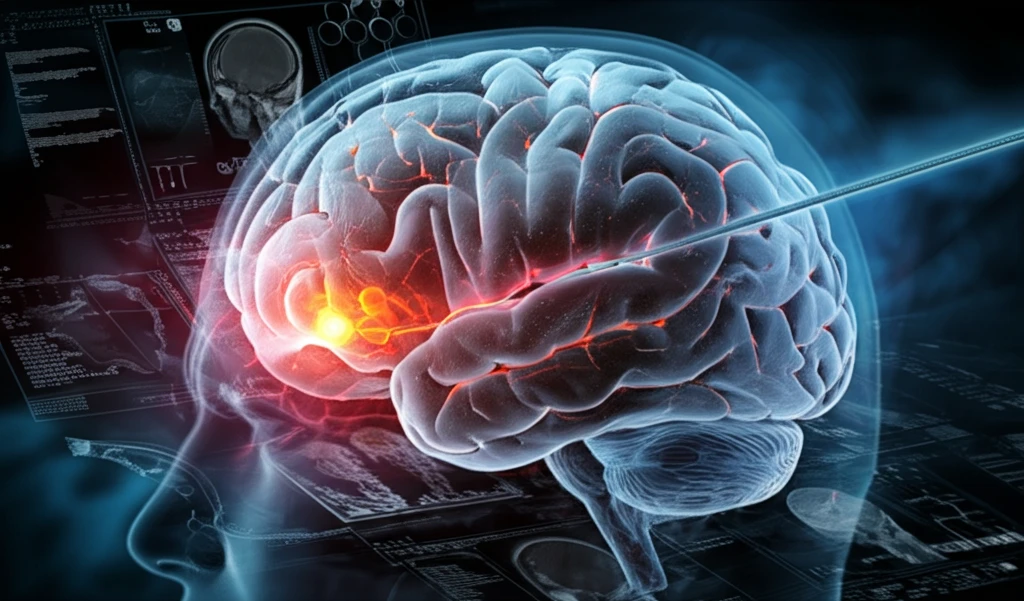
Stroke Breakthrough: Revolutionizing Treatment with Advanced Brain Imaging

"New research unveils how a tailored approach to stroke treatment, combining embolic occlusion techniques with sophisticated MRI, could transform patient outcomes."
Stroke remains a leading cause of disability and death worldwide, underscoring the urgent need for more effective treatments. Pre-clinical studies play a crucial role in the development of new therapies, often relying on animal models to mimic the complexities of human stroke. These models help researchers test the potential of thrombolytic agents and neuroprotective strategies before they reach clinical trials.
Traditionally, many stroke models have lacked the precision needed to accurately replicate the conditions seen in human patients. For example, some methods involve inducing blood clots in the common carotid artery, which can lead to unpredictable blockages in various parts of the brain. This makes it difficult to assess the true impact of potential treatments.
But what if we could create a more precise model, one that closely mirrors the way strokes occur in humans? A new study published in the Journal of Neuroscience Methods details a groundbreaking technique for selectively blocking the middle cerebral artery (MCA) in rabbits. This method, combined with advanced brain imaging, offers a promising platform for pre-clinical stroke research.
Stroke’s Ongoing Impact
Stroke remains a serious health concern because it can damage the brain and affect movement, communication, cognition, or independence. Its effects vary according to the area of the brain involved and how quickly treatment begins. Advanced brain imaging may help clinicians understand these changes, but its impact depends on timely access to appropriate care.
Established Care and Its Limits
Standard stroke care generally focuses on identifying the cause of the event, restoring or managing blood flow when appropriate, and supporting recovery. Treatment decisions must account for factors such as stroke type, timing, symptoms, and overall health. These approaches can be limited when diagnosis or treatment is delayed, when brain injury is extensive, or when specialized services are unavailable.
From Recognition to Modern Diagnosis
Stroke is a medical emergency caused either by blocked blood flow to the brain or by bleeding in the brain, according to Cleveland Clinic. Wikipedia reports that about 87% of strokes are ischemic, while the remainder are hemorrhagic, although hemorrhagic transformation can complicate classification. Modern public-health guidance, including the CDC’s May 19, 2026 signs-and-symptoms resource, emphasizes recognizing symptoms and seeking emergency treatment quickly.
How Does Selective Embolic Occlusion Work?
The key to this new technique lies in its precision. Researchers at UCLA developed a method using fluoroscopic-guided microcatheter placement to target the MCA. Here's a step-by-step breakdown:
- Using real-time imaging, the microcatheter is advanced into the internal carotid artery, past the posterior communicating artery.
- A cerebral angiogram confirms the catheter's position and ensures the anterior cerebral artery (ACA) and MCA are clear.
- A small, pre-formed blood clot is then injected through the microcatheter to create a blockage in the proximal MCA.
Understanding Stroke and the Brain
The American Stroke Association presents stroke education as a step-by-step explanation of its different types and effects on the brain. Its overview also includes prevention tools, linking understanding of stroke mechanisms with efforts to reduce risk. This broad framework provides context for evaluating how advanced brain imaging may support prevention, diagnosis, and treatment decisions.
Limits of Medical Innovation
Advanced imaging should not be treated as a complete solution to every stroke problem. Its value may be reduced when access is delayed, interpretation is uncertain, or imaging results cannot be converted into timely treatment. Clinical decisions must therefore continue to account for symptoms, timing, patient condition, and available expertise.
Imaging Within Stroke Care
Advanced brain imaging can add detail to stroke evaluation, but it does not replace the broader clinical process. Its usefulness must be considered alongside established assessment, treatment, and rehabilitation practices. In real-world care, the strongest approach is likely to be one that integrates imaging findings with clinical judgment and timely intervention.
The Future of Stroke Research
This innovative rabbit model offers a significant step forward in pre-clinical stroke research. By combining selective embolic occlusion with advanced MRI techniques, researchers can gain a more detailed understanding of how strokes evolve and how potential therapies might impact recovery. While further investigation is needed, this approach holds great promise for developing new and more effective treatments for stroke patients in the future.
Integrating Imaging and Care
Brain imaging is most meaningful when it helps clinicians make faster and better-informed decisions. It should be viewed as part of a coordinated stroke-care pathway rather than as an isolated breakthrough. The potential benefit lies in connecting clearer information about brain injury with appropriate treatment and follow-up.
The Next Phase of Stroke Treatment
Future progress may focus on making advanced imaging faster, more accessible, and more useful at the point of care. Research will also need to establish how imaging findings should guide treatment for different patients and stroke types. Any promising development will require careful validation before it becomes routine practice.
Access and Implementation
The benefits of advanced brain imaging depend not only on technical capability but also on healthcare infrastructure, trained professionals, and rapid referral systems. Unequal access could widen differences in stroke outcomes between communities. Implementation therefore requires attention to affordability, availability, workflow, and equitable delivery.
Patients Behind the Images
For people experiencing stroke, the practical value of imaging is measured by whether it supports faster diagnosis, effective treatment, and meaningful recovery. Families and caregivers also depend on clear communication about what scans show and what they mean for next steps. Technology can strengthen care, but compassion, coordination, and rehabilitation remain central to the human experience of recovery.