
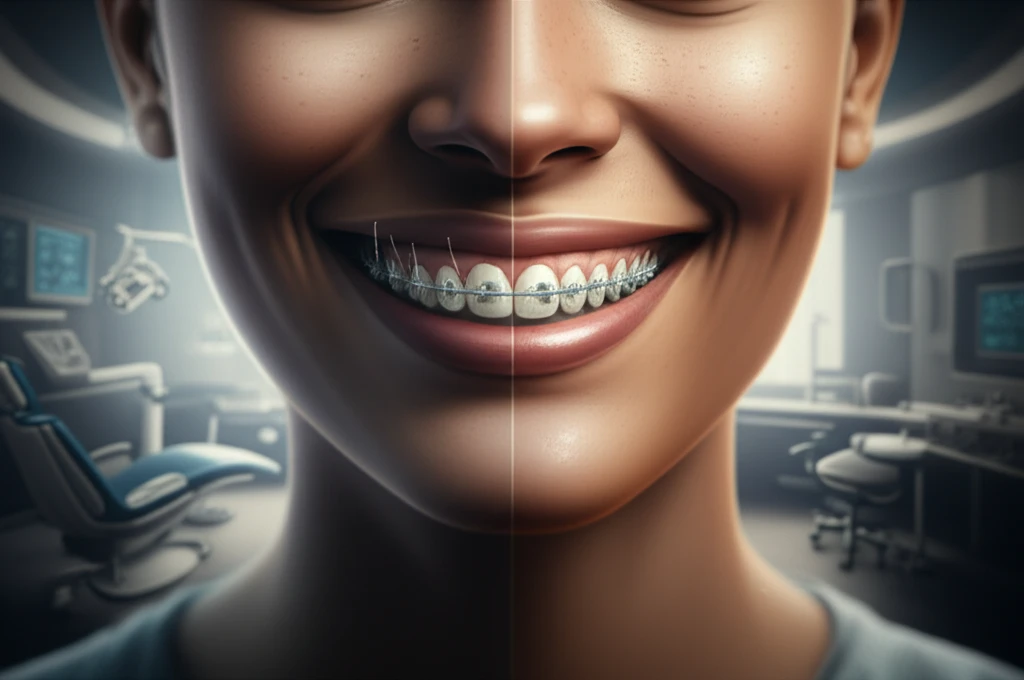
Smile Upgrade: Unveiling the Accuracy of Digital Dental Models for a Perfect Grin

"From Scans to Smiles: How digital models are reshaping the world of orthodontics, offering a glimpse into a future of precision and efficiency."
In the ever-evolving landscape of dental technology, the shift from traditional methods to digital solutions is undeniable. One of the most significant advancements is the use of digital dental models, which are created using intraoral scanners. These devices capture detailed images of a patient's teeth and gums, creating a 3D model that can be used for various orthodontic procedures.
This transition is not just about embracing new technology; it's about enhancing precision, improving efficiency, and ultimately, providing a better experience for patients. Digital models offer a level of accuracy that can be challenging to achieve with traditional methods, leading to more predictable and effective treatments. The promise of a perfect smile is now closer than ever, thanks to these innovative tools.
This article dives deep into the world of digital dental models, examining their accuracy and comparing them to traditional methods. We'll explore how these models are created, the benefits they offer, and what the future holds for this exciting technology. Prepare to discover how digital innovation is revolutionizing the path to a healthy, beautiful smile.
Current Impact
Digital dental models are increasingly presented as a useful tool in orthodontic diagnosis, treatment planning, and patient communication. Their potential impact lies in making dental structures easier to view and evaluate in three dimensions. However, the exact clinical benefits and accuracy can vary with the technology, workflow, and case being assessed.
Established Methods
Orthodontic assessment traditionally relies on clinical examination, dental records, imaging, and physical study models. Digital models offer a computer-based alternative that may improve viewing, storage, and measurement of dental structures. Their limitations may include dependence on scanning quality, software workflows, and the need for appropriate clinical interpretation.
From Plaster to Pixels
Dental study models have long been central to orthodontists' classification of malocclusion and development of treatment plans. The history of orthodontics spans roughly two millennia, while digital dental models emerged as a major technological development in the early 2000s. A 2004 introduction to digital models described how technological advances enabled three-dimensional models to be saved and viewed on computers, addressing several problems associated with conventional plaster models. Later reviews characterized digital 3D models as important advances used in orthodontic diagnosis, treatment planning, and appliance production, with accuracy reported as reaching or sometimes exceeding that of plaster models.
Digital Models vs. Traditional Methods: A Comparative Look
Traditional dental models rely on physical impressions, often made using materials like alginate. This process can be uncomfortable for patients and is prone to inaccuracies. Digital models, on the other hand, involve scanning the mouth with an intraoral scanner, which captures detailed images in real-time. These scans are then converted into 3D models using specialized software.
- Enhanced Precision: Digital models offer a higher degree of accuracy, reducing the margin of error in orthodontic treatments.
- Increased Efficiency: Digital scanning eliminates the need for physical impressions, saving time and reducing the number of appointments.
- Improved Patient Comfort: Digital scans are less invasive and more comfortable compared to traditional impression-taking methods.
- Better Communication: 3D models allow for enhanced visualization, improving communication between orthodontists and patients.
Current Research
Recent reviews are examining digital models across diagnosis, treatment procedures, retention, and outcome evaluation in orthodontics. One scoping review identified 87 studies for qualitative analysis by three reviewers, indicating a broad research base rather than a focus on a single clinical application. Another recent review emphasizes artificial-intelligence algorithms as tools for improving precision and reliability in orthodontic diagnostics and treatment strategies. Research published between 2018 and 2025 has also been synthesized to assess digital twin applications in diagnosis, treatment planning, and biomechanical simulation.
Limits and Uncertainty
Digital models and three-dimensional technology have become part of everyday orthodontic practice, but their necessity is not established for every case. A 2024 scoping review specifically reported that there is currently no consensus about which orthodontic procedures require a digital model. Reviews also describe combinations of digital models with CBCT images and rapid-prototyping techniques as promising, while indicating that some resulting treatment approaches are still regarded as future developments rather than universally established practice.
Digital Versus Manual Assessment
A 2025 comparison of orthodontic treatment evaluation methods concluded that manual and digital ABO-OGS assessments were clinically interchangeable. The study reported small statistical differences, while digital models produced reproducible results and 69% of cases met ABO-OGS passing criteria. These findings support digital models as a reliable alternative for orthodontic outcome evaluation, although the reported statistical differences show that the methods are not identical in every measurement.
The Future of Smiles: Embracing Digital Innovation
Digital dental models represent a significant leap forward in orthodontic care. They offer enhanced precision, increased efficiency, and improved patient experiences. As technology continues to evolve, we can expect even greater advancements in this field, leading to more predictable and effective treatments. The future of smiles is digital, and it's brighter than ever before.
What the Evidence Suggests
Taken together, digital dental models appear to be a practical addition to orthodontic assessment rather than a universal replacement for clinical judgment. Their value is likely greatest when three-dimensional visualization, repeatable measurements, or digital treatment planning are important. The evidence also suggests that clinicians should match the technology to the procedure and the patient's needs.
The Digital Orthodontic Future
Future orthodontic workflows may combine digital models with CBCT imaging to create a full three-dimensional virtual patient. This integrated view is intended to support thorough evaluation of skeletal and dental structures and help determine orthodontic needs. Digital orthodontic offices are also increasingly equipped to design and print appliances, including dental models, aligners, indirect-bonding trays, and occlusal splints. These developments point toward more comprehensive in-office digital production and treatment planning.
Implementation Challenges
Broader adoption of digital dental models may depend on access to suitable scanning, imaging, software, and fabrication equipment. It may also require consistent workflows and training so that measurements and treatment decisions remain dependable across clinical settings. These practical considerations can influence whether digital methods deliver their intended benefits.
Patients and Clinicians
Digital models can give clinicians a more accessible three-dimensional way to examine dental structures and plan care. For patients, clearer visual explanations may help make orthodontic goals and proposed treatments easier to understand. Even so, the technology supports rather than replaces the clinician's responsibility to interpret findings and tailor treatment to the individual.