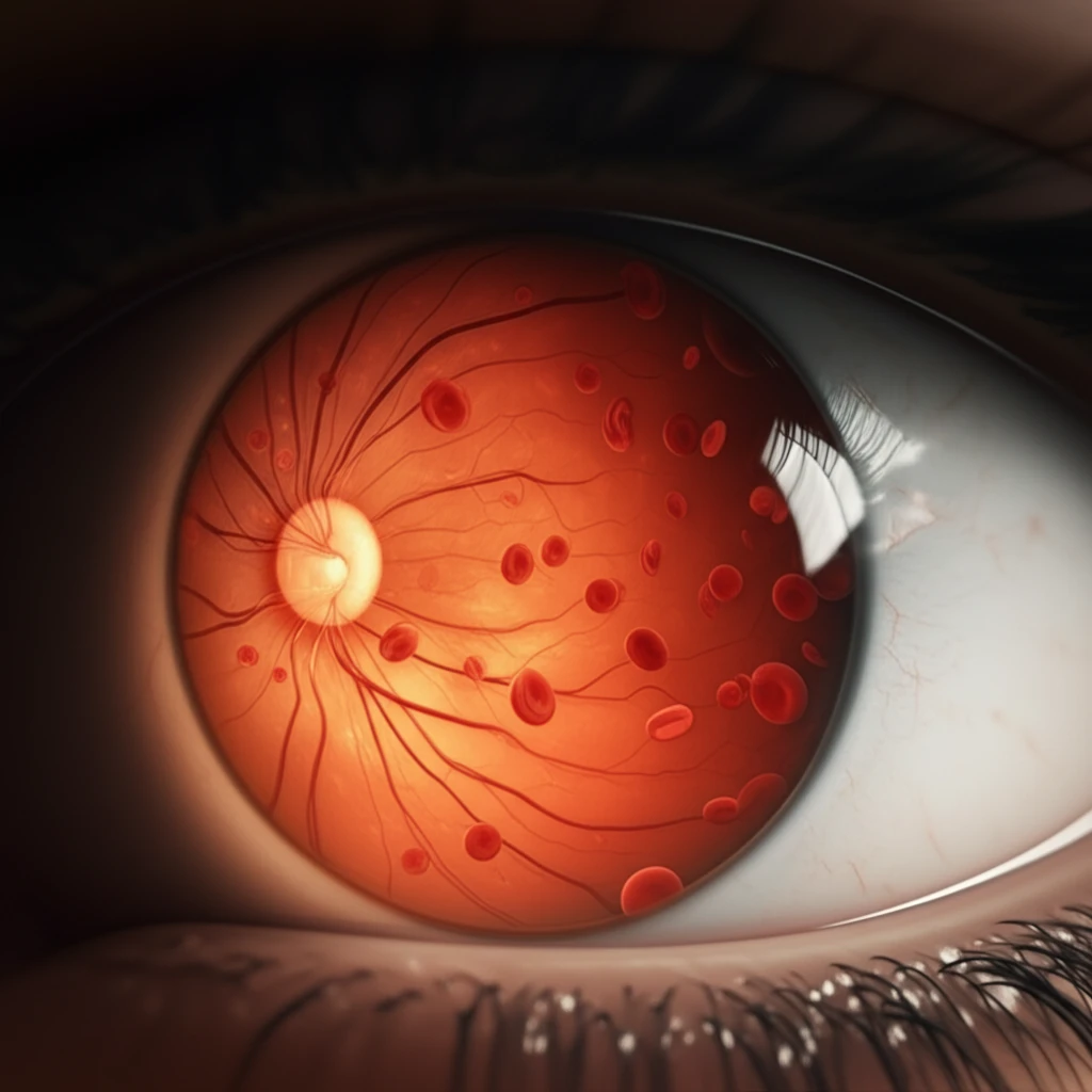
Stylized illustration of a human eye with intricate blood cell patterns representing sickle cell retinopathy.

Sickle Cell Retinopathy: Clearing Up the Confusion

"Navigating the Terminology and Diagnosis for Better Outcomes"


A recent study by Beral and associates shed light on the prevalence of retinopathy and maculopathy in patients with sickle cell disease (SCD), noting the absence of a direct link to hematological parameters, blood rheology, or genotype. While this research contributes valuable insights, it also highlights the need for clear and consistent terminology in the diagnosis and classification of these conditions.

The retina, a complex structure essential for vision, is divided into the posterior pole and the peripheral retina. Within the posterior pole lies the macula, a critical area for central vision. General terms like 'retinopathy' and 'maculopathy' simply refer to diseases affecting the retina and macula, respectively. However, when dealing with specific conditions like sickle cell retinopathy (SCR), a more detailed approach is crucial.

Since Goldberg's classification of SCR in 1971, it has served as the gold standard for diagnosing and classifying the disease through funduscopic examination. Macular involvement in SCD was first described in 1972, with traction maculopathy already recognized as related to proliferative SCR. Maculopathy is, therefore, an integral component of SCR, closely linked to peripheral vascular nonperfusion.

Retinopathy vs. Maculopathy: Defining the Terms in Sickle Cell Disease

Stylized illustration of a human eye with intricate blood cell patterns representing sickle cell retinopathy.

The study by Beral and colleagues employs the terms 'Retinopathy' and 'Maculopathy' seemingly as distinct entities. However, the diagnostic criteria and classifications used for each were not clearly defined. This raises questions about how 'Retinopathy' (peripheral vascular alterations) and 'Maculopathy' (macular ischemic impairment) were specifically assessed, and what stage of peripheral retinopathy and OCT findings were considered for diagnosis.

Fluorescein angiography (FA) is known to detect more advanced stages of peripheral SCR than clinical evaluation alone. Similarly, Optical Coherence Tomography (OCT) offers superior sensitivity in detecting sickle cell maculopathy compared to traditional fundus examination. Given these advancements, a comprehensive diagnostic approach is essential.

  • Fluorescein Angiography (FA): Detects peripheral SCR stages earlier.
  • Optical Coherence Tomography (OCT): More sensitive for detecting sickle cell maculopathy.
The researchers utilized a highly sensitive method (OCT) for detecting macular involvement but seemingly a less sensitive method for peripheral disease. Details regarding the specific methodologies employed in these tests are lacking in their article. Greater clarity regarding the study methodology, particularly concerning ophthalmological evaluations, would enhance understanding.

Moving Forward: Toward Enhanced Diagnostic Protocols

Clarifying the diagnostic criteria for retinopathy and maculopathy is crucial. Distinguishing between peripheral vascular changes and macular involvement is essential for accurate diagnosis and treatment planning.

The Dell'Arti et al. study highlights the potential of OCT technology in identifying systemic risk factors associated with sickle cell maculopathy, and suggests that chronic chelation therapy and elevated HbF levels may offer protective benefits. This underscores the importance of continued research into both diagnostic and therapeutic strategies.

By refining diagnostic approaches and incorporating advanced imaging techniques, healthcare professionals can improve early detection and management of SCR and maculopathy, ultimately preserving vision and enhancing the quality of life for individuals with sickle cell disease.

About this Article -

This article was crafted using a human-AI hybrid and collaborative approach. AI assisted our team with initial drafting, research insights, identifying key questions, and image generation. Our human editors guided topic selection, defined the angle, structured the content, ensured factual accuracy and relevance, refined the tone, and conducted thorough editing to deliver helpful, high-quality information.See our About page for more information.

This article is based on research published under:

DOI-LINK: 10.3233/ch-180462, Alternate LINK

Title: Terminologies Regarding Sickle Cell Retinopathy

Subject: Physiology (medical)

Journal: Clinical Hemorheology and Microcirculation

Publisher: IOS Press

Authors: Ricardo Luz Leitão Guerra, Mateus Abdalla Bastos, Cristina Salles

Published: 2019-06-27

Everything You Need To Know

1

What is Sickle cell retinopathy (SCR)?

Sickle cell retinopathy (SCR) is a condition affecting the retina, the light-sensitive tissue at the back of the eye, due to sickle cell disease (SCD). It encompasses a range of changes, including those in the peripheral retina and macula. The macula is a specific area within the posterior pole of the retina responsible for central vision. SCR can lead to vision problems if not diagnosed and managed effectively.

2

What is the difference between Sickle cell retinopathy (SCR) and maculopathy?

The distinction between Sickle cell retinopathy (SCR) and maculopathy is crucial for accurate diagnosis and effective treatment. SCR refers to the broader disease affecting the retina, whereas maculopathy specifically targets the macula. Maculopathy is often related to the changes happening in the peripheral retina. Understanding this differentiation helps doctors to tailor treatment based on the specific area and severity of the damage. It underscores the importance of detailed diagnostic approaches.

3

What is the significance of Goldberg's classification?

Goldberg's classification, established in 1971, remains the gold standard for diagnosing and classifying Sickle cell retinopathy (SCR) through funduscopic examination. This system provides a structured way to understand the progression and severity of the disease. While valuable, the evolution of diagnostic tools like Fluorescein Angiography (FA) and Optical Coherence Tomography (OCT) allows for earlier and more detailed detection of SCR and maculopathy, prompting the need for consistent terminology and diagnostic criteria.

4

How do Fluorescein Angiography (FA) and Optical Coherence Tomography (OCT) help in the diagnosis of Sickle cell retinopathy (SCR) and maculopathy?

Fluorescein Angiography (FA) and Optical Coherence Tomography (OCT) are advanced imaging techniques used in diagnosing Sickle cell retinopathy (SCR) and maculopathy. FA helps detect earlier stages of peripheral SCR by visualizing blood vessels in the retina. OCT offers superior sensitivity for detecting macular involvement compared to traditional fundus examination. The use of these tools improves diagnostic accuracy and enables timely intervention.

5

Why is clear terminology important when discussing Sickle cell retinopathy (SCR) and maculopathy?

The study by Beral and associates underscores the importance of clear and consistent terminology in the diagnosis and classification of Sickle cell retinopathy (SCR). It highlights how unclear diagnostic criteria for conditions like retinopathy and maculopathy can lead to ambiguities in understanding disease prevalence and progression. More detailed methods in future studies will help refine our understanding of SCR and macular involvement in patients with sickle cell disease.

Newsletter Subscribe

Subscribe to get the latest articles and insights directly in your inbox.