Surreal digital illustration symbolizing combined kV/MV CBCT imaging advancements in medical diagnostics.

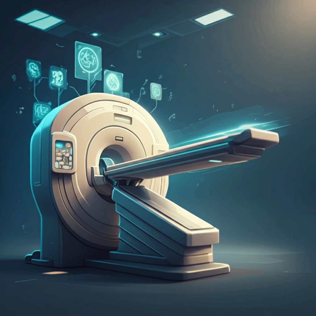
Revolutionizing Medical Imaging: Can Combined kV/MV CBCT Scans Reduce Scan Time and Metal Artifacts?

"A deep dive into a new study exploring the potential of combined kilovoltage/megavoltage cone-beam computed tomography (kV/MV CBCT) with a high-DQE MV detector to enhance image quality and streamline medical imaging processes."


In the ever-evolving landscape of medical imaging, precision and efficiency are paramount. State-of-the-art medical linear accelerators are now commonly equipped with two imaging systems: an electronic portal imaging device (EPID) used with the treatment beam and an orthogonal kilovoltage (kV) system. These systems offer complementary information, improving localization accuracy and treatment quality.

Cone-beam computed tomography (CBCT) or kV planar imaging is typically used for patient setup, beam gating, and beam delivery verification, while the MV system is more commonly used for treatment quality assurance and exit dosimetry. Recent research explores the potential of combined kilovoltage/megavoltage (kV/MV) imaging as an avenue for innovation, focusing on reducing scan times and minimizing metal artifacts—common challenges in radiotherapy and diagnostics.

A recent study investigates the use of combined kV/MV CBCT imaging with a high Detective Quantum Efficiency (DQE) MV detector. The goal is to determine if this advanced setup can generate acceptable quality pre-treatment CBCT images at clinically acceptable dose levels. By addressing limitations related to scan time and image distortion, this research opens new possibilities for enhancing image-guided radiotherapy applications.

How Does Combined kV/MV CBCT Imaging Work?

Surreal digital illustration symbolizing combined kV/MV CBCT imaging advancements in medical diagnostics.

The study, conducted using a Truebeam system, combined data from both 6MV and 100kVp projections. The MV data was acquired using a prototype EPID containing two scintillators: a standard copper-gadolinium oxysulfide (Cu-GOS) screen and a prototype focused cadmium tungstate (CWO) pixelated “strip.” The kV data was acquired using a standard onboard imager. Image quality was then evaluated using phantoms—an 18-cm diameter electron density phantom and a 20-cm diameter Catphan phantom—analyzing contrast and resolution.

To simulate metal artifact reduction (MAR), researchers replaced two CIRS phantom inserts with steel rods. They based reconstruction methods on combining MV and kV data into a single sinogram, using mostly kV raw data and replacing rays corrupted by metal with MV data. For scan time reduction (STR), projections from partially overlapping kV and MV acquisitions were combined, and the resultant images were compared against MV-only and kV-only reconstructions.

  • Scan Time Reduction (STR): Projections from partially overlapping 105° kV and MV acquisitions were combined to create a complete data set, potentially achievable in just 18 seconds.
  • Metal Artifact Reduction (MAR): The reconstruction process utilized primarily kV raw data, with MV data selectively replacing rays corrupted by metal, thus reducing distortions.
  • Dose Levels: The total absorbed dose for MAR was approximately 0.7 cGy, while the STR combined acquisition resulted in about 2.5 cGy.
The results indicated that the high-DQE MV detector significantly improved image quality for both STR and MAR applications. In combined kV/CWO STR reconstruction, all contrast inserts were visible, whereas only two were detectable in the kV/Cu-GOS image due to high noise levels. Similarly, in kV-MV MAR reconstructions, streaking artifacts were substantially reduced, making all inserts clearly visible in the kV/CWO image.

The Future of Medical Imaging

This study confirms that a high-DQE MV detector can be effectively applied to generate high-quality combined kV/MV images for SRT and MAR, using clinically acceptable doses. By significantly reducing scan times and minimizing artifacts caused by metal implants, this technology promises to improve diagnostic accuracy and streamline radiotherapy planning. As medical imaging technology continues to advance, innovations like combined kV/MV CBCT imaging will play a crucial role in shaping the future of healthcare.

About this Article -

This article was crafted using a human-AI hybrid and collaborative approach. AI assisted our team with initial drafting, research insights, identifying key questions, and image generation. Our human editors guided topic selection, defined the angle, structured the content, ensured factual accuracy and relevance, refined the tone, and conducted thorough editing to deliver helpful, high-quality information.See our About page for more information.

Everything You Need To Know

1

What are the primary benefits of using combined kilovoltage/megavoltage (kV/MV) cone-beam computed tomography (CBCT) in medical imaging?

The key advantages of combined kV/MV CBCT imaging include significantly reduced scan times and the minimization of metal artifacts. By integrating data from both kV and MV projections and using a high Detective Quantum Efficiency (DQE) MV detector, this approach enhances image quality, leading to quicker diagnoses and improved accuracy in radiotherapy planning. The method reduces distortions caused by metal implants which helps to produce clearer images compared to using kV or MV alone.

2

How does the combined kV/MV CBCT imaging technique specifically address the issue of metal artifacts in medical images?

To reduce metal artifacts, the reconstruction process in combined kV/MV CBCT imaging primarily uses kV raw data, but strategically replaces rays corrupted by metal with MV data. This method is based on the principle that MV data is less susceptible to metal artifacts due to the higher energy of the megavoltage beam. By combining these data sets, distortions and streaking artifacts caused by metal implants are significantly reduced, resulting in clearer visualization of the regions surrounding the metal. The prototype EPID is an integral part of this.

3

What role does the high Detective Quantum Efficiency (DQE) MV detector play in enhancing the performance of combined kV/MV CBCT imaging?

The high DQE MV detector is critical because it improves image quality for both scan time reduction (STR) and metal artifact reduction (MAR) applications. In STR, it allows for visualization of contrast inserts that would otherwise be undetectable due to high noise levels when using standard detectors like the copper-gadolinium oxysulfide (Cu-GOS) screen. Similarly, in MAR, the high DQE MV detector enables clear visualization of inserts by substantially reducing streaking artifacts, which are significant factors in obtaining high-quality images. The prototype focused cadmium tungstate (CWO) pixelated strip enhances the images obtained.

4

Can you explain how combined kV/MV CBCT imaging achieves a reduction in scan time, and what are the typical dose levels associated with this technique?

Scan time reduction (STR) in combined kV/MV CBCT is achieved by combining projections from partially overlapping kV and MV acquisitions to create a complete dataset. For instance, projections from 105° kV and MV acquisitions can be combined, potentially completing a scan in approximately 18 seconds. The total absorbed dose for scan time reduction using this combined acquisition is about 2.5 cGy. This efficient data acquisition process minimizes the overall time required for imaging without compromising image quality, using a Truebeam system, contributing to a streamlined workflow in medical settings.

5

What are the implications of using combined kV/MV CBCT imaging with a high-DQE MV detector for the future of medical imaging and radiotherapy planning?

The use of combined kV/MV CBCT imaging with a high-DQE MV detector signals a significant advancement in medical imaging and radiotherapy planning. By effectively reducing scan times and minimizing metal artifacts, this technology enhances diagnostic accuracy, leading to more precise and efficient radiotherapy planning. This also enables the generation of high-quality combined kV/MV images at clinically acceptable dose levels. As medical imaging technology continues to evolve, innovations like combined kV/MV CBCT imaging will play a crucial role in improving patient outcomes and shaping the future of healthcare, contributing to more effective and personalized treatment strategies.

Newsletter Subscribe

Subscribe to get the latest articles and insights directly in your inbox.