
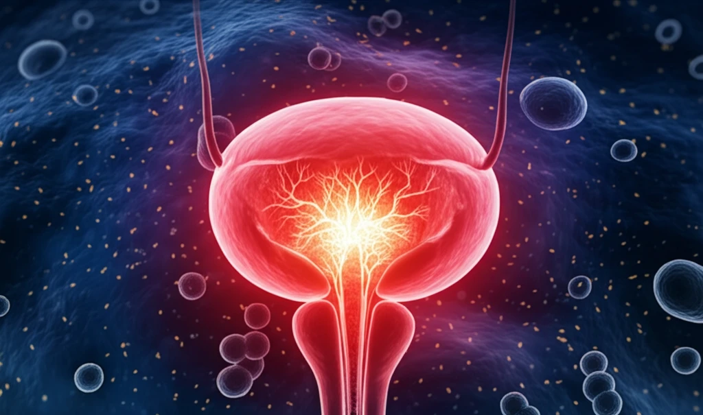
Pressure Points: Unlocking Bladder Health Through Cell Science

"Could Understanding Cellular Responses to Pressure Revolutionize Urological Treatments?"
Bladder reconstructive surgery has become a common procedure for addressing functional and anatomical issues within the bladder. Enterocystoplasty, while helpful, carries risks like infection, metabolic disorders, and malignancies. Tissue engineering is now a promising alternative, and key to its success is understanding how to maintain healthy bladder cells in the lab, specifically bladder smooth muscle cells and urothelial cells (UCs).
Urothelial cells (UCs) line the urinary tract, forming a crucial barrier that protects against water, ion, solute, and pathogen penetration. These cells, arranged in layers (basal, intermediate, and umbrella cells), rely on uroplakins—specialized cell membrane proteins—for their function. Uroplakins make up about 90% of umbrella cells and are vital for the barrier function of the UCs.
However, when UCs are isolated and grown in the lab, they tend to lose their specialized characteristics and functions. Therefore, encouraging and maintaining uroplakin expression is essential for effectively engineering bladder tissue. Emerging research highlights the importance of mechanical cues in cell differentiation. This raises an important question: Can cyclic hydrodynamic pressure (simulating the bladder's natural filling and emptying) help maintain uroplakin expression in UCs?
Uroplakins in Bladder Diagnosis
Uroplakin II (UPII) is a transmembrane protein relatively specific to urothelial cells, though its expression may be reduced in high-grade tumors. UPII antibodies are frequently used to assess whether a primary or metastatic lesion originates from the urothelium. Uroplakin 1B (Upk1b) helps stabilize bladder-lining epithelial cells and may prevent them from rupturing during bladder distension. A separate study investigated plasma and urine uroplakin IIIa (UPIIIa) levels in bladder cancer, including their relationship to chemical carcinogen exposure, DNA damage, and detoxification ability in smokers.
Markers and Subtype Classification
Bladder cancer is classified into basal and luminal molecular subtype categories, which have treatment and prognostic implications. A two-marker immunohistochemistry approach using GATA3 and CK5/6 has been reported to provide over 80% reliability for predicting these subtypes. A 2022 study evaluated uroplakin II as a single marker for distinguishing luminal from basal tumors. Uroplakin III expression has also been examined in transitional cell carcinoma, while the available source describes the uroplakin promoter as a tool for bladder cancer gene therapy but says the protein’s role in the bladder remained unknown.
How Does Cyclic Hydrostatic Pressure Impact Uroplakin Expression?
Researchers investigated the impact of cyclic hydrostatic pressure on uroplakin expression in human UCs and the role of extracellular regulated protein kinases 1/2 (ERK1/2) in this process. They subjected human UCs to varying levels of cyclic hydrodynamic pressure (simulating bladder cycles) and then analyzed uroplakin expression using real-time PCR and western blot techniques. They also used an ERK1/2 inhibitor to determine the involvement of ERK1/2 signaling.
- Cyclic hydrostatic pressure, specifically at 200 cm H2O, can significantly enhance uroplakin expression in human UCs.
- ERK1/2 signaling is involved in the pressure-induced uroplakin expression.
- Inhibiting ERK1/2 reduces the pressure-induced uroplakin expression.
Pressure, Mechanotransduction, and Drug Delivery
An Arduino-controlled, 3D-printed millifluidic platform was introduced to emulate clinical pressure waveforms in cell culture using 96-well plates. The system is described as a high-throughput hydrostatic pressure exposure platform. A review identifies Piezo channels, integrins, YAP/TAZ, and TRPV4 as key effectors responding to mechanical stimulation. Separately, research on Piezo1-mediated drug delivery frames the bladder as operating in a complex biomechanical stress environment.
Expression Can Change, and Defects Matter
In control bladder, uroplakin III expression appeared strongly along the epithelium, but a reported experiment found a significant decrease or loss in intact bladder mucosa 12 hours after cyclophosphamide injection. This illustrates that uroplakin expression can vary under an experimentally induced bladder injury condition. Other work cited in the source material links uroplakin defects to a compromised urothelial permeability barrier, vesicoureteral reflux, and abnormal voiding patterns. Uroplakins form plaques on the apical membranes of umbrella cells, where they contribute to the barrier that limits urine passage across the urothelium.
Uroplakin Antibody Detection
The supplied comparison source describes uroplakin III as a highly specific but moderately sensitive immunohistochemical marker for primary and metastatic urothelial carcinomas. It also cites a 2014 report of a newly developed uroplakin II antibody with increased sensitivity in urothelial carcinoma of the bladder. A separate 2022 source notes that little was known about uroplakin II protein expression in urothelial carcinoma, possibly because a commercially available uroplakin II antibody was absent. Together, these reports highlight differences in the stated sensitivity of markers and the availability of antibodies across the cited material.
Why This Matters for Bladder Health
This study sheds light on how mechanical forces influence uroplakin expression in UCs, with implications for tissue engineering and treating urinary tract diseases. By identifying the optimal pressure conditions (200 cm H2O) and the involvement of the ERK1/2 signaling pathway, researchers have opened new avenues for enhancing uroplakin expression in engineered bladder tissues. Uroplakins are not only markers of UC differentiation but also play roles in bladder cancer diagnostics and interstitial cystitis. Understanding how to regulate their expression could lead to new therapeutic strategies for these conditions.
Uroplakin Expression and Tumor Progression
A 2023 study set out to evaluate the prognostic role of uroplakin expression in urothelial carcinomas and describes loss of Upk1a and Upk1b expression as linked to stage progression. Upk1a and Upk1b have also been identified as diagnostic markers for distinguishing urothelial carcinomas from other solid tumor entities. Upk1b is described as helping stabilize bladder-lining epithelial cells and prevent rupture during bladder distension. These findings position uroplakins as relevant both to tissue identification and to bladder epithelial structure, while the supplied excerpts do not provide specific prognostic results.
Human Biopsies and Diagnostic Markers
A 2022 study examined human bladder biopsies to investigate urothelial responses to urinary tract infection, uroplakin expression, and healing after cystoscopy with electrofulguration (CEF) for antibiotic-recalcitrant recurrent UTI. The biopsies included regions described as “no cystitis” and “cystitis” from women with antibiotic-recalcitrant disease. Separately, a 2026 study aimed to assess GATA3, UPII, and p40 individually and as a combined immunohistochemistry panel in urothelial carcinoma, including correlations with tumor grade and invasion status. The supplied excerpt describes the study aims but does not report its results.