
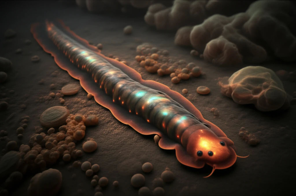
Planarian Regeneration: Unlocking the Secrets of Wound Healing with ODC-like RNAi

"New research reveals how ODC-like proteins in planarians could revolutionize our understanding of tissue repair and regenerative medicine."
For centuries, the remarkable ability of certain creatures to regenerate lost limbs or heal severe wounds has captivated scientists and the public alike. Among these extraordinary organisms, planarians—tiny flatworms—stand out due to their seemingly limitless regenerative capacity. Unlike humans, who respond to injury with scarring, planarians can regrow entire body parts, even from small fragments. This incredible feat has made them a focal point for regenerative medicine research.
Recent studies have increasingly pointed to polyamines, naturally occurring compounds involved in cell growth and differentiation, as critical players in various signaling pathways. However, scientists are still working to fully understand how these molecules orchestrate the complex processes of cell growth and tissue repair. A key enzyme in polyamine production is ornithine decarboxylase (ODC), which is tightly regulated by intricate molecular machinery. Further complicating the picture, multiple ODC-like proteins exist, and some appear to lack the ability to catalyze reactions, suggesting alternative roles in the ODC regulatory pathway.
Now, a groundbreaking study published in Scientific Reports sheds light on the function of ODC-like proteins in planarians, offering new perspectives on wound healing and regenerative processes. Researchers investigated six planarian ODCs (ODC1-6) and discovered that silencing specific ODC homologs led to a unique wound response phenotype, independent of putrescine—a common polyamine. This discovery challenges existing assumptions about the role of polyamines in planarian regeneration and opens up new avenues for exploring regenerative medicine.
Planarian Regeneration and Translational Potential
Planarian regeneration involves all tissue types and is regulated by distinct molecular factors and cellular signaling pathways, with neoblasts playing vital roles in regeneration and morphology maintenance. A 2026 bibliometric analysis reported that, despite more than two centuries of research, the field's knowledge landscape remains fragmented, limiting interdisciplinary integration and translational progress. In experimental models, planarian extracellular vesicles significantly accelerated tissue regeneration and wound closure while improving re-epithelialization and barrier integrity compared with controls.
Decoding ODC's Role in Planarian Regeneration
The research team began by identifying and characterizing six ODC homologs in the planarian species Dugesia japonica. Through detailed bioinformatic analysis, they found that five of these homologs (ODC2-6) contained substitutions in key amino acids essential for enzymatic activity. This suggested that these ODC-like proteins might not function as typical decarboxylases.
Emerging Research Directions
Recent work has linked microtubule-associated protein 1B to cytoskeletal activity and neurotransmitter release at the planarian nervous-system level. Research using artificially altered gravity evaluated molecular responses in intact and regenerating planarians and reported that cerium oxide nanoparticles exerted a protective antioxidant action. Another study used high doses of 5-fluorouracil to deplete Dugesia japonica stem cells and identified a transiently resistant population of lineage-committed stem cells.
Limits of the ODC-like RNAi Interpretation
The reported putrescine-independent wound-response phenotype produced by ODC-like RNAi complicates a simple interpretation based only on putrescine depletion. Among the planarian ODC paralogs examined, only DjODC1 in D. japonica and Smed-odc-A in S. mediterranea lacked substitutions in all 18 key amino acids. The source therefore identifies these two paralogs as the candidates most consistent with fully functional ornithine decarboxylase activity, while leaving open the possibility that other ODC-like proteins have altered functions.
Comparing Regenerative Variables
A study of DjODC5 RNAi reported that particular morphological changes appeared in 100% of intact animals, or 30 of 30 specimens. Separate work on lymphocyte activation examined differential metabolism of radioactive putrescine and spermidine after B-cell mitogen LPS exposure, providing a comparison point for how polyamine metabolism can vary by molecule and biological context. Other sources frame planarians as useful for investigating human tissue repair and report that regeneration effectiveness can be affected by environmental solutions.
Implications for Regenerative Medicine
This research sheds light on the complexities of planarian regeneration and highlights the potential for ODC-like proteins to contribute to wound healing in unexpected ways. By identifying novel functions for these proteins, scientists can gain a deeper understanding of the molecular mechanisms that drive tissue repair. This knowledge could one day be harnessed to develop new therapies for wound healing and regenerative medicine, potentially leading to treatments that minimize scarring and promote tissue regeneration in humans.
Equinox and Regenerative Positioning
The planarian wound epidermis gene equinox is expressed in the wound epidermis. The reported study identifies equinox as essential for initiating positional-information regeneration and blastema formation. This places wound-epidermis gene activity near the beginning of the process that establishes regenerative organization.
From ODC-like RNAi to Coordinated Repair
Research reported by Stanford indicates that distant wounds help planarians heal and that whole-body coordination is crucial for wound healing and subsequent tissue regeneration. The ODC-like RNAi findings likewise highlight unexpected functions for ODC-like proteins beyond a straightforward putrescine-dependent wound response. Together, these findings point toward future work connecting local molecular perturbations with coordinated, organism-wide repair mechanisms.
Long-Range Wound Signaling
In planarians, injury induces extracellular signal-regulated kinase activity waves that travel at speeds reported to be 10 to 100 times faster than those in other systems. Longitudinal body-wall muscles function as pathways for propagating wound signals and disseminating systemic responses. Inhibiting Erk propagation and distal wound responses blocked regeneration, while a second amputation of distal tissue within a narrow time window rescued regeneration.