
Microtubule Magic: How Cell Structure Proteins Fine-Tune The Architecture of Life

"Unlocking the Secrets of EB Proteins: A Deep Dive into Cellular Dynamics and Spatial Organization"
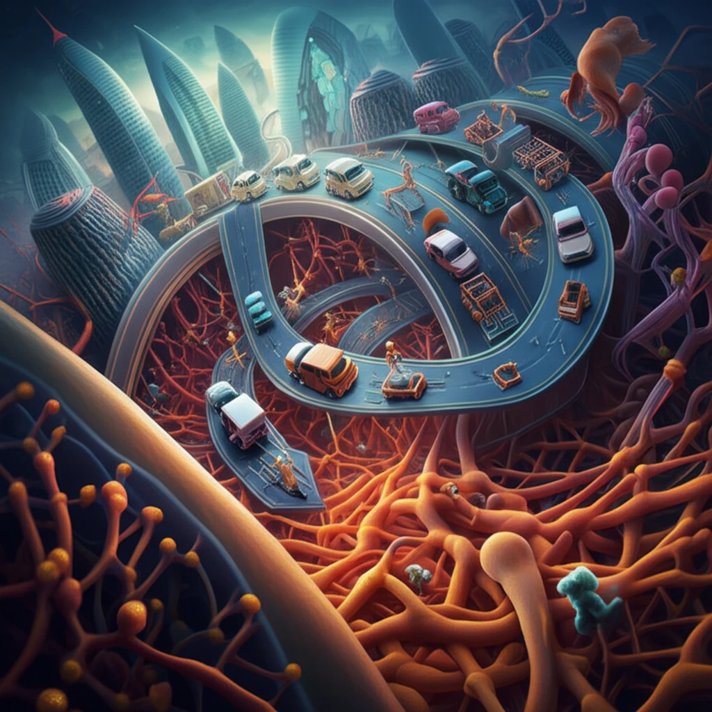
In the bustling metropolis of our cells, microtubules act as crucial highways, guiding essential cargo and providing structural support. These dynamic polymers are constantly assembling and disassembling, a process tightly regulated by a cast of proteins, most notably the End-Binding (EB) family. These proteins track the tips of growing microtubules, acting as hubs for a network of other proteins that coordinate cell division, cell movement, and overall cell shape. Understanding how EB proteins function is critical to unlocking the secrets of cellular life.
Like skilled construction workers, EB proteins ensure that microtubules are built correctly and positioned precisely. In mammalian cells, three main EB proteins—EB1, EB2, and EB3—each play unique roles. While EB1 is universally expressed, EB2 and EB3 are more selective, appearing in specific cell types and under certain conditions. This begs the question: what makes these EB proteins different, and how do their distinct properties contribute to the diverse functions of our cells?
New research has peeled back the layers of this complex system, revealing that the spatial positioning of EB proteins at microtubule tips involves distinct nucleotide-dependent binding properties. By reconstituting EB1, EB2, and EB3 activity in vitro, scientists have discovered that each protein prefers to bind to slightly different configurations of tubulin, the building blocks of microtubules. These subtle preferences dictate where each EB protein localizes on the microtubule tip, influencing the entire architecture of the cell.
Microtubules and Cellular Architecture
Microtubules are protein-based components of the cytoskeleton that help organize the interior of cells. Their dynamic structure allows cells to maintain shape and coordinate essential internal activities. The precise scale of their impact varies by cell type and biological context, so broad conclusions should be made cautiously.
Tubulin-Based Construction
The standard structural model describes microtubules as hollow tubes formed when alpha- and beta-tubulin combine into dimers, polymerize into protofilaments, and associate laterally. The most common arrangement consists of 13 protofilaments forming the tubular wall. This framework explains the basic architecture of microtubules, although descriptions of their broader cellular roles can vary by source and context.
Building the Modern Model
The modern understanding of microtubules has developed through increasingly detailed studies of cell structure and protein assembly. Researchers have gradually connected their visible filament architecture with the behavior of tubulin subunits. Because no subsection-specific historical sources were provided here, this overview remains general rather than assigning particular discoveries or dates.
Why Does EB Protein Placement Matter? The Nucleotide Code
Microtubules are made from tubulin, which binds to GTP. When tubulin is incorporated into the microtubule structure, the GTP is hydrolysed (broken down by water) into GDP. The balance between GTP and GDP tubulin at the microtubule tip determines whether the microtubule grows or shrinks.
- EB1 and EB3: These proteins are similar in their preference and bind strongly to the GTP analogues and prefer regions rich in GTP-tubulin. They rapidly exchange at the microtubule tip, indicating a dynamic interaction.
- EB2: EB2 behaves differently; it binds best to microtubule lattices containing a mix of different nucleotides. It uniquely prefers lattices with a 1:1 ratio of different nucleotides, demonstrating a specific affinity.
An Active Research Frontier
Current microtubule research continues to examine how these structures assemble, reorganize, and interact with other cellular components. Studies increasingly treat microtubules as dynamic systems rather than static support rods. Without sources specific to this subsection, the pace and implications of recent findings cannot be stated more precisely.
Limits of Simplified Models
A simplified account of microtubules may overlook differences among cell types, assembly conditions, and interacting proteins. Structural descriptions alone do not necessarily capture the full complexity of cellular behavior. In the absence of subsection-specific sources, these limitations should be treated as general cautions rather than evidence for a particular disputed claim.
Comparing Cellular Structures
Microtubules are one part of the broader cytoskeletal framework that gives cells internal organization. Their properties and effects are best understood in relation to other structural systems and to the needs of particular cells. No source material was supplied here to support a detailed comparison or rank one system as universally superior.
The Big Picture: How EB Proteins Shape Our Cells
By understanding the unique binding preferences of EB1, EB2, and EB3, we gain insight into how cells orchestrate a wide range of functions. From cell division to intracellular transport, the precise spatial organization of these proteins is crucial. This research highlights the importance of subtle molecular differences in achieving complex cellular behavior. With further exploration, scientists can continue to decode the intricate mechanisms that govern our cells, paving the way for future advances in medicine and biotechnology.
A Dynamic Cellular Framework
Microtubules can be viewed as both structural elements and adaptable organizers within the cell. Their importance lies in how their architecture supports changing cellular arrangements rather than merely providing rigid support. This synthesis is intentionally broad because no expert commentary was supplied for this subsection.
Questions Ahead
Future work will likely seek clearer explanations of how microtubule organization is controlled across different cellular environments. Progress may depend on linking molecular-scale assembly with whole-cell behavior. Specific predictions about technologies, discoveries, or timelines would require sources that were not provided here.
Structure Within a Larger System
Microtubules operate within complex cells rather than as isolated structures. Their behavior is therefore connected to broader questions about coordination, regulation, and the interaction of many biological systems. Without subsection-specific evidence, the systemic challenges can only be described in general terms.
Why Cellular Structure Matters
Understanding microtubules can help place cellular organization within the larger study of life and health. Their biology may matter to people through research, education, and future medical investigation, but the practical consequences depend on the specific biological or clinical context. No sources were provided here to support a particular therapy, disease connection, or measurable human outcome.