
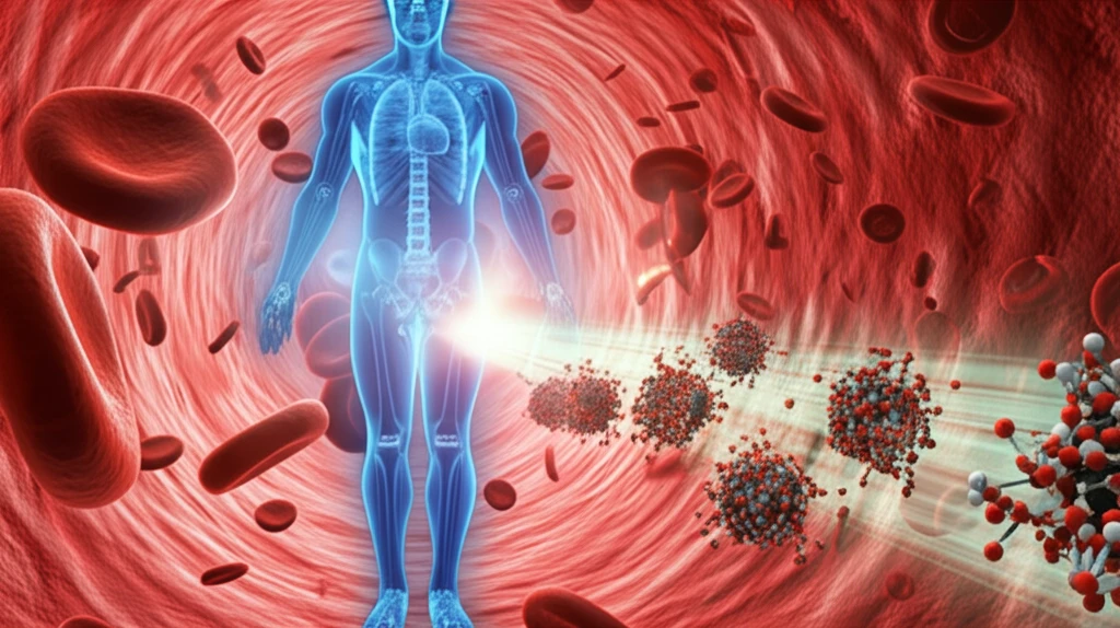
Melanin Nanoparticles: The Future of Medical Imaging?

"Explore how melanin nanoparticles chelated with barium ions offer a safer, naturally-derived alternative to traditional X-ray contrast agents."
In modern medicine, contrast enhancement agents are essential for improving the clarity and detail of medical imaging techniques. X-ray computed tomography (CT) relies on these agents to visualize internal structures. However, the commercially available contrast agents often fall short of ideal, prompting researchers to explore safer and more effective alternatives.
Currently, barium sulfate suspensions and iodinated molecules are commonly used for gastrointestinal and vascular CT imaging, respectively. These iodinated contrast agents have drawbacks, including short blood circulation time, rapid renal clearance (leading to a brief imaging window), potential toxicity at high concentrations, and a lack of specificity in how they distribute within the body.
In recent years, nanoparticulate systems, especially organic and inorganic nanoparticles, have garnered significant attention as potential X-ray contrast agents. Their ability to track cells and target specific tissues makes them attractive for advanced imaging applications. Nanoparticles containing elements with high atomic numbers excel at absorbing X-rays, resulting in enhanced image contrast. However, concerns about the poor degradability and potential toxicity of inorganic nanoparticles have limited their widespread use.
Melanin Nanoparticles in Biomedical Imaging
Melanin is a naturally occurring biopolymer found in living organisms that has emerged as a promising material for biomedical imaging applications. It is prized for its biocompatibility and biodegradability, along with properties including scavenging abilities, metal chelation, and electronic conductance. Melanin-based nanoparticles can function as contrast agents themselves or serve as nanocarriers for imaging, controlled drug release, and therapeutic applications. Researchers have synthesized melanin-like nanoparticles (MNPs) with good dispersion stability in biological media, achieved through surface modifications such as thiol-terminated methoxy-poly(ethylene glycol) coatings.
Current Contrast Agents and Their Limitations
Current CT imaging relies on conventional contrast agents such as barium sulfate suspensions and iodinated molecules, which have known limitations in clinical practice. Melanin-like nanoparticles have made progress as photoacoustic (PA) contrast agents for in vivo imaging in animal studies. However, researchers note that the construction of degradable or metabolizable melanin-based nanoparticles is necessary, especially for future clinical applications. Advances in photon-counting detector CT (PCD-CT) hardware and software, including image processing techniques for noise reduction, are considered crucial for driving novel applications and facilitating clinical translation of new contrast agents.
Foundations of Melanin Research in Medicine
Melanin research in medicine has evolved from studying this pigment primarily as a biological coloring agent to recognizing its potential as a multifunctional biomaterial. Early investigations focused on understanding melanin's natural role in organisms, including its protective properties against radiation and oxidative stress. The transition toward medical imaging applications built upon discoveries about melanin's broad-spectrum light absorption and its ability to interact with various imaging modalities. While the field has progressed significantly, much of the foundational work on melanin-based contrast agents remains in preclinical stages.
Why Melanin Nanoparticles Could Revolutionize CT Imaging
Scientists are actively seeking nano-platforms to overcome the challenges associated with existing contrast agents. Liposomes, micelles, and protein nanoparticles are being developed as efficient platforms for delivering drugs and contrast agents. However, liposomes and micelles are delicate systems that require complex handling and formulation processes. Protein nanoparticles need cross-linkers and specific binding ligands, making their synthesis intricate and complicated.
- Natural Chelation: Melanin's inherent ability to chelate metal ions is vital for regulating their amounts in biosystems.
- Enhanced Chelation at Nanoscale: The chelating ability of melanin increases at the nanoscale due to the high surface-to-volume ratio of melanin nanoparticles (MNPs).
- Ideal Candidate: Considering melanin's strong chelating ability, melanin NPs are ideal as a new nano-platform in bio-imaging.
Recent Advances in Nanoparticle Contrast Agents
Recent reviews have highlighted developments in nanoparticle contrast agents for photon-counting computed tomography (PCCT), with research published in early 2025 examining translational opportunities and key considerations for clinical translation. Additionally, a comprehensive 2026 review systematically summarized advances in CT contrast agent research, focusing on novel molecular design and synthesis strategies, optimization of pharmacokinetics and imaging performance, expansion of clinical application fields, and safety management including strategies to address adverse reactions. These studies emphasize that the advent of PCCT technology creates new possibilities for nanoparticle-based imaging that were not feasible with conventional CT systems.
Challenges in Melanin Nanoparticle Development
Early research on melanin nanoparticles as contrast agents for optoacoustic tomography, published in 2015, demonstrated the synthesis and characterization of melanin-like nanoparticles (MNPs). While the work established proof-of-concept for using MNPs as novel contrast agents, the research also underscored the challenges inherent in translating these materials from laboratory synthesis to clinical application. The difficulty in achieving consistent results and the need for extensive optimization represent ongoing hurdles in the field.
Melanin vs. Traditional Imaging Agents
Comparative analysis of melanin-based nanoparticles against traditional contrast agents reveals both advantages and limitations. Melanin offers inherent biocompatibility and biodegradability, properties that synthetic agents typically lack. However, traditional agents have established clinical track records and well-understood pharmacokinetics that melanin nanoparticles have yet to match. The field continues to evaluate where melanin nanoparticles can provide unique benefits rather than simply replicating existing capabilities.
The Promise of Melanin in Medical Imaging
Melanin nanoparticles chelated with barium ions hold great promise as a CT contrast agent, offering similar or even slightly higher X-ray attenuation than commercially available iodinated agents. This innovative approach leverages the natural properties of melanin to create a biocompatible and biodegradable contrast agent, paving the way for safer and more effective medical imaging techniques. With further research and development, melanin-based contrast agents could become a transformative tool in diagnostic imaging, enhancing our ability to visualize and understand the complexities of the human body.
Expert Perspectives on Melanin Imaging
The scientific community recognizes melanin nanoparticles as a promising but still-developing platform for medical imaging. Experts note that while the material's natural properties are advantageous, significant work remains before clinical adoption. The consensus appears to be that melanin-based agents will likely complement rather than replace existing contrast technologies in the near term. Continued interdisciplinary collaboration between materials scientists, clinicians, and regulatory experts will be essential for advancing the field.
Emerging Directions in Melanin Research
Future research directions for melanin nanoparticles in imaging include developing more sophisticated surface modification techniques to enhance targeting capabilities and imaging performance. The integration of melanin nanoparticles with other diagnostic modalities, such as dual-modal MRI/fluorescence imaging, represents an active area of investigation. Regulatory pathways and manufacturing scalability will need to be addressed as the technology matures toward potential clinical applications.
Technical Barriers to Clinical Translation
Despite melanin's potential as an MRI contrast agent, most melanin-like nanoparticles face limitations due to their large size and poor solubility. Developing melanin-like contrast agents with excellent solubility and high stability is considered important for future clinical application. Research has explored innovative solutions such as silica-coated metal chelating-melanin nanoparticles, which function as dual-modal contrast agents for MRI and fluorescent imaging while also serving cancer therapeutics. These approaches demonstrate how surface engineering can address fundamental material limitations.
Biomedical Applications of Melanin Nanoparticles
Melanin-based nanoparticles have emerged as a versatile platform in biomedicine owing to their intrinsic biocompatibility, broadband light absorption, and radical scavenging properties. These characteristics make them particularly attractive for applications where biological safety is paramount. The natural origin of melanin may also help address patient concerns about synthetic materials being introduced into their bodies. As research progresses, the potential for melanin nanoparticles to improve diagnostic capabilities while minimizing side effects continues to generate interest across the medical community.