Illustration of a healthy liver with glowing vines contrasted with shadowy nodules, representing liver health and potential risks.

Liver Nodules Got You Worried? A Simple Guide to Understanding Your Risks

"Decoding Hypovascular Nodules: What They Mean for Your Liver Health and How to Stay Proactive"


Hepatocellular carcinoma (HCC), a common and deadly cancer, often lurks silently. Early detection is crucial, and advancements in imaging techniques, like contrast-enhanced MRI, have improved our ability to spot potential problems. However, these scans can sometimes reveal small, hypovascular nodules—areas that appear to have reduced blood flow—leaving patients and doctors wondering about the next steps.

One such advancement is the use of Gadolinium-ethoxybenzyl-diethylenetriamine pentaacetic acid (Gd-EOB-DTPA) in MRI. This liver-specific contrast agent helps visualize how well liver cells are functioning. When nodules appear hypointense (darker) during the hepatobiliary phase of a Gd-EOB-DTPA-enhanced MRI, it indicates a potential issue with liver cell function. But what does this really mean for your health?

This article explores a recent study investigating these hypovascular nodules. We'll break down the key findings, offering a clear understanding of the factors that might predict progression to HCC and what you can do to stay informed and proactive about your liver health.

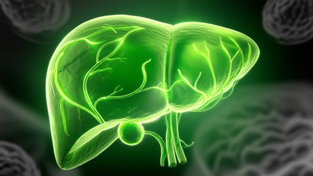
What are Hypovascular Nodules and Why Do They Matter?

Illustration of a healthy liver with glowing vines contrasted with shadowy nodules, representing liver health and potential risks.

Hypovascular nodules are areas in the liver that show reduced blood flow compared to the surrounding tissue. They are often detected during imaging tests like MRI or CT scans, particularly when contrast agents are used. These nodules appear darker or less enhanced than the rest of the liver because they don't absorb as much of the contrast agent, indicating a difference in blood supply and cell function.

The concern with hypovascular nodules is that they can sometimes be an early sign of hepatocellular carcinoma (HCC). However, not all hypovascular nodules turn into cancer. Many remain stable or even disappear over time. The challenge for doctors is to determine which nodules are likely to progress to HCC and which are not.

  • Potential Precursors to Cancer: Some hypovascular nodules can develop into HCC.
  • Benign Conditions: Many nodules are non-cancerous and remain stable.
  • Diagnostic Dilemma: Determining which nodules will progress is challenging.
Recent research has focused on identifying factors that can help predict the likelihood of a hypovascular nodule progressing to HCC. By understanding these factors, doctors can better monitor patients and make informed decisions about treatment.

The Takeaway: Staying Informed and Proactive About Your Liver Health

While the study highlights the difficulty in predicting which hypovascular nodules will progress to HCC based solely on initial background factors, it also underscores the importance of diligent monitoring. If you have been diagnosed with hypovascular nodules, regular follow-up appointments and imaging tests are essential for tracking any changes and detecting potential problems early. By staying informed and working closely with your healthcare team, you can take proactive steps to protect your liver health and overall well-being.

About this Article -

This article was crafted using a human-AI hybrid and collaborative approach. AI assisted our team with initial drafting, research insights, identifying key questions, and image generation. Our human editors guided topic selection, defined the angle, structured the content, ensured factual accuracy and relevance, refined the tone, and conducted thorough editing to deliver helpful, high-quality information.See our About page for more information.

This article is based on research published under:

DOI-LINK: 10.2169/internalmedicine.8801-16, Alternate LINK

Title: Progression Of Hepatic Hypovascular Nodules With Hypointensity In The Hepatobiliary Phase Of Gd-Eob-Dtpa-Enhanced Mri In Hepatocellular Carcinoma Cases

Subject: General Medicine

Journal: Internal Medicine

Publisher: Japanese Society of Internal Medicine

Authors: Tsukasa Saitoh, Shuichi Sato, Tomotaka Yazaki, Hiroshi Tobita, Tatsuya Miyake, Shunji Ishihara, Takashi Katsube, Hajime Kitagaki, Yoshikazu Kinoshita

Published: 2018-01-01

Everything You Need To Know

1

What are hypovascular nodules in the liver, and why are they a cause for concern?

Hypovascular nodules are areas within the liver that exhibit reduced blood flow compared to the surrounding liver tissue. They are often detected through imaging techniques like MRI or CT scans, particularly when contrast agents are used. The concern arises because these nodules can potentially be an early indicator of hepatocellular carcinoma (HCC), a type of liver cancer. However, it's important to note that not all hypovascular nodules progress to cancer; many remain stable or even disappear over time. Therefore, determining which nodules are likely to develop into HCC is a key challenge for doctors.

2

How does a contrast-enhanced MRI using Gadolinium-ethoxybenzyl-diethylenetriamine pentaacetic acid (Gd-EOB-DTPA) help in evaluating liver nodules?

Gadolinium-ethoxybenzyl-diethylenetriamine pentaacetic acid (Gd-EOB-DTPA) is a liver-specific contrast agent used in MRI to visualize how well liver cells are functioning. During the hepatobiliary phase of a Gd-EOB-DTPA-enhanced MRI, if nodules appear hypointense (darker), it suggests a potential issue with liver cell function. This contrast helps in better characterization of liver nodules compared to traditional MRI contrasts, aiding in the assessment of their malignant potential. Further, the intensity of the nodule during the hepatobiliary phase provides crucial information about the functionality of the hepatocytes within the nodule, which is vital for diagnosis.

3

If hypovascular nodules are detected, what steps should I take to proactively manage my liver health?

If you have been diagnosed with hypovascular nodules, diligent monitoring is essential. Regular follow-up appointments with your healthcare team and scheduled imaging tests are crucial for tracking any changes in the nodules and detecting potential problems early. Discuss with your doctor about lifestyle modifications, such as avoiding alcohol and maintaining a healthy weight, which can help reduce the risk of liver disease progression. It's also vital to inform your doctor about any family history of liver cancer or existing liver conditions, as these factors can influence the monitoring and management strategy for hypovascular nodules.

4

What are some factors that might predict whether a hypovascular nodule will progress to hepatocellular carcinoma (HCC)?

While predicting the progression of hypovascular nodules to hepatocellular carcinoma (HCC) based solely on initial background factors remains a challenge, certain characteristics and risk factors are considered. These may include the size and growth rate of the nodule, the presence of certain imaging features, and underlying liver conditions such as cirrhosis or hepatitis. Regular monitoring and imaging can help identify changes in the nodule that might suggest progression. Further research is ongoing to identify more specific biomarkers and imaging characteristics that can improve the accuracy of predicting HCC development from hypovascular nodules.

5

Given that not all hypovascular nodules turn into cancer, what are some other possible explanations for their appearance on liver scans?

While hypovascular nodules can be an early sign of hepatocellular carcinoma (HCC), it's important to remember that many are benign. These non-cancerous nodules can result from various conditions, including focal nodular hyperplasia (FNH), liver hemangiomas, or regenerative nodules in the context of cirrhosis. Additionally, some inflammatory conditions or prior liver injuries can lead to the formation of hypovascular nodules. The key to differentiating between benign and potentially malignant nodules lies in comprehensive evaluation, including detailed imaging analysis and, in some cases, biopsy to determine the specific nature of the nodule.

Newsletter Subscribe

Subscribe to get the latest articles and insights directly in your inbox.