
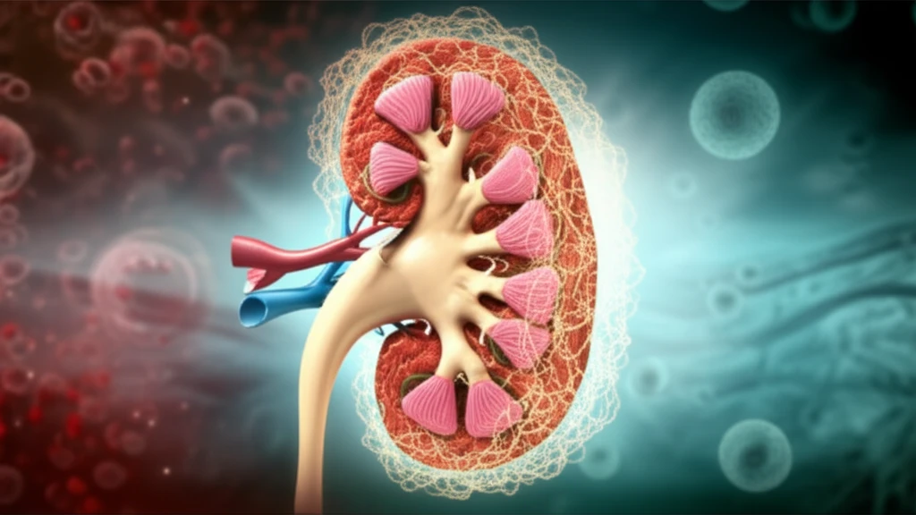
Kidney Protection: Can We Prevent Damage from Hypoxia?

"New research highlights how targeting a specific protein could shield your kidneys from the harmful effects of oxygen deprivation."
Acute kidney injury (AKI) is a serious condition that involves a rapid decline in kidney function, often requiring immediate medical attention. One of the major causes of AKI is ischemia-hypoxia, where the kidneys don't receive enough oxygen. This lack of oxygen can trigger a cascade of harmful processes, leading to significant damage.
The body has a natural response to low oxygen levels, primarily managed by a protein called hypoxia-inducible factor-1α (HIF-1α). HIF-1α helps the body adapt, but it's tightly controlled by another group of proteins called prolyl hydroxylase domain (PHD) proteins. Think of PHDs as the 'oxygen sensors' – they regulate how much HIF-1α is active. Among these, PHD2 is considered the most critical sensor.
New research explores whether inhibiting PHD2 could protect the kidneys during hypoxic events. By examining human kidney cells and a mouse model of ischemia-reperfusion injury (a process that mimics the oxygen deprivation and subsequent restoration seen in AKI), scientists are uncovering promising insights into kidney protection.
Why Kidney Hypoxia Matters
Kidney hypoxia is increasingly recognized as a potential contributor to kidney injury and chronic disease. Reduced oxygen availability may disrupt normal renal function and contribute to progressive damage, although the scale and clinical impact vary across conditions. Preventing or limiting hypoxic injury therefore remains an important research goal.
Detection and Current Strategies
Research on renal hypoxia uses multiple detection methods to assess oxygen availability and hypoxia-related signaling in kidney tissue. Reviews emphasize that each method has advantages and disadvantages, so careful interpretation is needed when evaluating renal hypoxia and potential treatments for hypoxia-associated CKD. Current mechanistic work also examines how oxygen metabolism, CKD progression, and HIF activation relate to kidney protection, including effects indexed by VEGF, HO-1, Epo, and GLUT1.
From Hypoxia Hypothesis to Debate
A major line of research proposed that renal tissue hypoxia helps drive CKD and may represent a final common pathway to end-stage renal failure. This view encouraged therapeutic strategies designed to increase kidney oxygenation and prompted investigation of oxygen supply, oxygen metabolism, and factors that produce hypoxia in CKD. Later reviews, including work on the HIF pathway and HIF prolyl hydroxylase inhibitors, have examined evidence both supporting and challenging the hypoxia hypothesis.
PHD2's Role: How Does Inhibiting It Help?
The study initially confirmed that when kidney cells (specifically, HK-2 tubular epithelial cells) were exposed to a chemical mimicking hypoxia (cobalt chloride, or CoCl₂), both PHD2 and HIF-1α levels increased. Interestingly, this exposure also led to increased cell death and activation of autophagy, a cellular 'self-eating' process that, while sometimes protective, can also contribute to cell damage.
- Autophagy Inhibition: Using a drug called 3-MA, they blocked autophagy. This protected the kidney cells from CoCl₂-induced damage, suggesting that in this scenario, autophagy was harmful.
- PHD2 Knockdown: They used siRNA to specifically reduce PHD2 levels in the cells. This also protected the cells from CoCl₂, potentially by increasing HIF-1α expression.
- HIF-1α Reversal: When they then reduced HIF-1α levels in the PHD2-inhibited cells, the protective effect disappeared, highlighting HIF-1α's crucial role.
- In Vivo Studies: Mice pretreated with a PHD inhibitor (L-mimosine) experienced less kidney damage from ischemia-reperfusion injury. The drug reduced cell death and inflammation in the injured kidneys.
Hypoxia-Responsive Kidney Research
A July 17, 2025 study reported that a hypoxia-responsive tRNA-derived small RNA, tRNA-Asp-GTC-3′tDR, is generated in human embryonic kidney cells and depends critically on the endonuclease angiogenin. The researchers validated its biogenesis using ultrasensitive Northern blotting and an engineered fluorescent reporter. Related reporting described this RNA as activating mechanisms that protect kidney cells from hypoxia-related damage, while reviews continue to examine hypoxia signaling and remaining questions about clinical use.
Why the Hypoxia Model Is Not Simple
The CKD literature links hypoxia with altered oxygen supply, oxygen metabolism, renal injury, and CKD-related complications. However, the available review evidence does not establish that every effect of hypoxia is uniformly harmful or that correcting oxygen levels alone will prevent disease progression. This complexity is one reason hypoxia-based interventions require careful evaluation rather than being treated as a single, universally sufficient solution.
A Conditional Picture
Research on kidney hypoxia presents a conditional rather than uniformly positive or negative picture. Some work treats hypoxia as a driver of renal injury, while other investigations examine hypoxia signaling as a possible route to cellular protection. Comparisons therefore need to account for the disease setting, the biological pathway being measured, and whether the evidence comes from experimental models or clinical research.
The Future of Kidney Protection: What Does This Mean?
This research offers a promising new avenue for protecting kidneys from the damaging effects of hypoxia. By targeting PHD2, scientists may be able to develop treatments that reduce the severity of AKI and other kidney-related conditions. The study suggests that activating HIF, via inhibiting PHD, may be a useful therapeutic strategy in ischemic renal injury.
A Nuanced Interpretation
The evidence suggests that kidney hypoxia is biologically important but cannot be interpreted in isolation from oxygen metabolism and the broader CKD process. Protection may depend on selectively activating adaptive pathways rather than simply increasing oxygen exposure. Because the available evidence includes both damaging and potentially protective effects, conclusions should remain appropriately cautious.
Targeting HIF-1α
Future kidney-protection strategies are being developed around regulation of HIF-1α through synthetic drugs and natural active compounds. A January 12, 2026 review reported that HIF prolyl hydroxylase inhibitors can stabilize HIF-1α, activate the HIF-1α/VEGFA/VEGFR1 signaling axis, and increase SOD2. The broader hypoxia market is also described as expanding amid rising disease burden, greater awareness, and investment in vascular and neurological therapeutics, although market activity does not itself establish clinical effectiveness.
Consequences Beyond Oxygen Levels
Kidneys are particularly vulnerable to disturbances in oxygen availability, and kidney hypoxia can disrupt essential physiological functions. According to the reviewed source, these consequences may extend to both acute and chronic kidney injury regardless of the underlying cause. This makes kidney protection a systemic challenge involving the cause of hypoxia, the kidney's response, and the timing of intervention.
Diabetes and Kidney Risk
Renal hypoxia is being studied as part of the pathogenesis of diabetic kidney disease. A review of the field considers chronic hypoxia, cellular mechanisms, and evidence from both animal and human studies, while also presenting hypoxia as a possible therapeutic target. More recent analysis emphasizes that hypoxia's role in CKD is nuanced, suggesting that real-world treatment decisions must distinguish harmful disease processes from potentially adaptive hypoxia responses.
While these findings are encouraging, it's important to remember that this is early-stage research. Further studies are needed to fully understand the mechanisms involved and to determine the safety and effectiveness of PHD2 inhibitors in humans. The interplay between autophagy, apoptosis, and inflammation is complex, and more research is needed to fully elucidate the signaling pathways involved.
For individuals at risk of AKI (e.g., those undergoing major surgery, with chronic kidney disease, or with conditions like diabetes), these findings highlight the importance of managing risk factors and seeking prompt medical attention if kidney problems arise. As research progresses, new strategies for kidney protection may become available, offering hope for improved outcomes.