
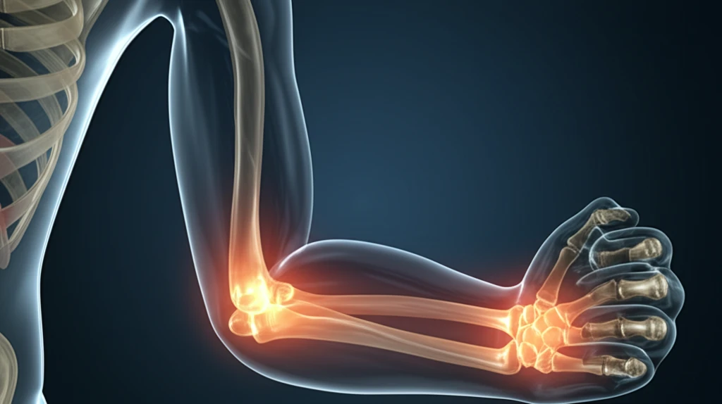
Is That Fracture Just a Little Bit Broken? Why Pediatric Elbow Injuries Need a Second Look

"MRI Scans Offer a Safer Way To Determine the Extent of Non-Displaced Humeral Condyle Fractures in Kids, Potentially Reducing Need for Surgery"
Elbow fractures are among the most common injuries in childhood, often resulting from playground tumbles, sports mishaps, or accidental falls. One particular type of elbow fracture, known as a non-displaced lateral humeral condyle fracture, presents a unique challenge for doctors. In these fractures, the bone may be cracked, but the pieces haven't shifted out of alignment by much, making it difficult to fully evaluate the extent of damage with traditional X-rays.
Traditionally, doctors have relied on X-rays to diagnose and manage these fractures. However, the elbow joint in children is largely made of cartilage, which doesn't show up well on X-rays. This limitation can lead to underestimating the severity of the fracture, potentially resulting in complications such as secondary displacement (where the fracture shifts later on), the development of non-unions (where the bone fails to heal properly), or the need for more invasive surgical interventions. For parents, this uncertainty can be unsettling, as they navigate the best course of treatment for their child's injury.
Now, a new approach using Magnetic Resonance Imaging (MRI) is offering a more precise way to assess these fractures. A recent study published in the Revue de Chirurgie Orthopédique et Traumatologique explores the use of MRI scans to get a clearer picture of the fracture's extent, potentially guiding treatment decisions and improving outcomes for young patients. Let's delve into how MRI is changing the game for pediatric elbow fractures and what it means for your child's care.
The Scale of Pediatric Elbow Fractures
Pediatric elbow fractures represent a significant portion of childhood musculoskeletal injuries. Medial epicondylar fractures alone are the third most common elbow fracture in children, typically seen in boys between the ages of 9 and 14. Large-scale retrospective data has catalogued thousands of cases, with one study of 4,607 pediatric elbow fractures revealing that lateral condyle fractures constituted 492 of those cases, and roughly 6.1% presented with concomitant elbow dislocation. These figures underscore that pediatric elbow injuries are neither rare nor trivial.
Why Standard Assessment Falls Short
The initial approach to a pediatric elbow injury follows a standard musculoskeletal assessment framework: a thorough history, physical examination, and radiographic imaging. However, the Radiology Assistant notes that elbow fractures are among the most common in children yet remain among the most difficult to assess, owing to the changing anatomy of the growing skeleton and the subtlety of some fracture patterns. Type I and Type II supracondylar fractures, in particular, can be clinically subtle and easy to miss, even when using established landmarks such as the anterior humeral line. This combination of high incidence and diagnostic difficulty is precisely why these injuries demand careful, repeated evaluation.
Building the Knowledge Base Over Decades
Clinicians have long recognized that certain pediatric elbow fracture types carry disproportionately high risks. Lateral condyle fractures, established as the second most common pediatric elbow fracture, are characterized by a higher risk of nonunion, malunion, and avascular necrosis than other fracture types. Supracondylar fractures, meanwhile, account for roughly 60% of all pediatric elbow fractures and are most commonly caused by a fall on an outstretched hand. Standard treatment has historically involved splinting or casting for approximately four weeks, reflecting decades of clinical refinement in managing these injuries.
MRI: A Clearer Picture of the Break
The study, led by C. Thévenin-Lemoine and colleagues, investigated the use of MRI in children with non-displaced lateral humeral condyle fractures. The core question was whether MRI could accurately determine if a fracture was complete (extending all the way through the bone) or incomplete (only partially through). This distinction is crucial because complete fractures are more likely to become displaced and may require surgery, while incomplete fractures can often be treated with a cast alone.
- MRI could indeed differentiate between complete and incomplete fractures in most cases.
- Out of 25 interpretable MRI scans, 17 showed incomplete fractures, while 8 revealed complete fractures.
- In two cases where the MRI revealed complete fractures, secondary displacement occurred, leading to surgical intervention.
- There was no correlation between the child's age and the completeness of the fracture, highlighting the importance of imaging for all ages.
AI, Radiology, and New Diagnostic Frontiers
Recent research is increasingly exploring how artificial intelligence can assist in detecting pediatric elbow fractures, reflecting a broader push to address diagnostic challenges. A radiographic review published in 2023 emphasizes that thorough knowledge of common pediatric elbow injuries is essential to their correct assessment and management, reinforcing that expertise remains foundational even as tools evolve. Meanwhile, a BMJ review highlights that medial epicondyle fractures account for approximately 10% of pediatric elbow injuries, adding to the epidemiological picture clinicians rely on. These developments suggest the field is moving toward integrating computational tools with established clinical knowledge.
The Problem of Unappreciated Fractures
A persistent concern in pediatric orthopedics is that certain elbow fractures remain chronically underappreciated by clinicians. Dr. Kaye Wilkins has specifically drawn attention to pediatric elbow fractures that are frequently overlooked or underestimated in clinical settings, highlighting a gap between available knowledge and real-world diagnostic practice. This underscores that the challenge is not purely one of medical knowledge but of clinical vigilance and awareness. Even well-established fracture patterns can be missed when they do not present in their textbook form.
Supracondylar Fractures vs. Other Elbow Injuries
Supracondylar humerus fractures stand out as the single most common fracture in the elbow area and the second most common of all pediatric fractures overall, surpassed only by distal radius fractures. They typically appear between the ages of five and seven, and unlike some other pediatric elbow fractures, no significant gender difference has been observed. This demographic profile contrasts with medial epicondylar fractures, which skew toward older boys aged 9 to 14, illustrating that different fracture types follow distinct epidemiological patterns. Understanding these differences is critical for guiding age-appropriate diagnostic and treatment strategies.
Making Informed Decisions for Your Child’s Elbow
The use of MRI in evaluating pediatric elbow fractures represents a significant advancement in orthopedic care. For parents, this means a more accurate diagnosis, a more tailored treatment plan, and potentially, a less invasive approach to healing their child’s broken elbow. While X-rays remain a valuable tool, MRI offers an additional layer of information that can lead to better outcomes and greater peace of mind. Discuss with your healthcare provider whether an MRI is right for your child's specific injury, and together, make an informed decision about their care.
The Core Takeaway
Pediatric elbow injuries sit at a challenging intersection of high prevalence and diagnostic subtlety. The growing skeleton introduces anatomical complexity that can obscure fractures from initial evaluation, while certain fracture types carry meaningful long-term risks if missed or undertreated. Continued investment in both clinical education and emerging diagnostic technologies appears essential to closing the gap between what is known and what is consistently applied in practice.
Changing Incidence and Evolving Patterns
The COVID-19 pandemic appears to have altered pediatric elbow fracture presentation patterns, with one study identifying 477 patients presenting with elbow fractures during the pandemic period, compared against a baseline drawn from a 2007–2017 database. This shift suggests that changes in childhood activity levels, healthcare access, or both can meaningfully affect injury epidemiology. As artificial intelligence tools mature, they may help clinicians better track and respond to such evolving patterns while improving individual diagnostic accuracy.
The Complexity of the Growing Skeleton
Pediatric elbow trauma remains one of the more challenging areas of musculoskeletal medicine because of the inherently complex nature of the growing skeleton. Radiographic landmarks shift as children develop, and chronic injuries can complicate the acute presentation picture. Addressing these challenges requires sustained investment in specialized pediatric imaging expertise and ongoing research into both anatomy and injury mechanisms.
What These Numbers Mean for Families
Behind the statistics are children whose elbow injuries can affect daily function, participation in sports, and long-term skeletal development if not caught early. A missed or delayed diagnosis can mean additional procedures, longer recovery times, and lasting consequences for a growing child. Ensuring that caregivers and clinicians alike understand the importance of thorough evaluation—especially when initial X-rays appear ambiguous—remains a critical part of reducing preventable harm.