
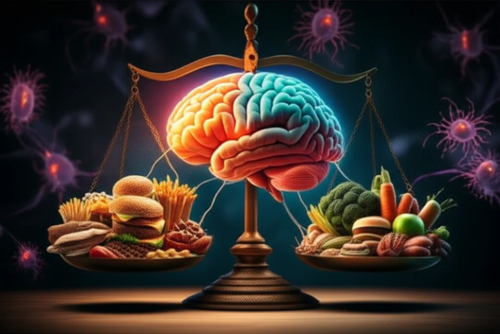
Is Obesity Really a Brain Game? How Hypothalamic Inflammation Changes Everything

"Uncover the surprising link between brain inflammation and weight gain, and what you can do about it."
For years, the battle against obesity has been framed as a simple equation: calories in versus calories out. While diet and exercise undoubtedly play crucial roles, mounting evidence suggests a more complex picture. Scientists are beginning to understand that obesity may have as much to do with what's happening in your brain as it does with what's on your plate.
Specifically, research is focusing on the mediobasal hypothalamus (MBH), a key brain region involved in controlling hunger, energy expenditure, and metabolism. When this area becomes inflamed, it can disrupt the delicate balance that regulates body weight, making it harder to lose weight and easier to gain it.
This inflammation often involves microglia, the brain's resident immune cells. When triggered by factors like a high-fat diet, these cells become activated, leading to a cascade of events that can alter brain function and contribute to obesity. This article explores the fascinating connection between hypothalamic inflammation, microglial activation, and weight gain, offering insights into potential new strategies for managing obesity.
Obesity and Brain Inflammation
Obesity is associated with chronic, low-grade systemic inflammation and insulin resistance, with growing evidence that the brain is a particularly vulnerable target organ. Reviews describe how obesity-related peripheral inflammation can affect central nervous system physiology and generate neuroinflammation. The blood-brain barrier may help mediate these effects, while hypothalamic circuits involved in whole-body energy homeostasis and cognitive function may also be affected.
Current Clinical Approach
Standard approaches to obesity generally combine dietary, physical-activity, behavioral, and medical strategies. However, a brain-centered explanation suggests that these approaches may not fully capture the biological effects of hypothalamic inflammation, insulin resistance, and altered energy regulation. Because the evidence spans interacting metabolic, inflammatory, and neurological processes, conclusions about treatment limitations should remain appropriately cautious.
From Body to Brain
Research on obesity has increasingly expanded from peripheral metabolism to communication between adipose tissue, the blood-brain barrier, and the central nervous system. This shift has supported the view that hypothalamic regulation and neuroinflammation may be relevant to whole-body energy balance. The historical importance of these developments lies in connecting metabolic disease with brain physiology, although the field continues to evolve.
The Hypothalamus: Your Brain's Control Center for Weight Management
The mediobasal hypothalamus (MBH) is like your brain's mission control for energy balance. It constantly monitors your body's energy status and coordinates responses to keep things running smoothly. Within the MBH, the arcuate nucleus (ARC) acts as a first responder, directly sensing signals from the body, such as hormones like leptin and insulin.
- Proopiomelanocortin (POMC) and Cocaine- and Amphetamine-Regulated Transcript (CART) neurons: These neurons are activated after a meal, suppressing appetite and increasing energy expenditure.
- Neuropeptide Y (NPY) and Agouti-Related Protein (AgRP) neurons: These neurons kick in when you're fasting, stimulating hunger and reducing energy expenditure.
The Emerging Obesity-Brain Axis
Recent reviews describe obesity-related disruption within the central nervous system, including increased oxidative stress, chronic inflammation, abnormal protein accumulation, mitochondrial dysfunction, hormonal imbalances, insulin resistance, and blood-brain barrier impairment. The literature also identifies obesity as an emerging risk factor for cognitive impairment and dementia. Therapeutic reviews further describe inflammation beginning in adipose tissue and propagating through peripheral organs via immune responses or macrophage activation.
Limits of the Inflammation Model
The inflammation-centered model remains a proposed explanation rather than a complete account of obesity-related brain disorders. One review reports that adipose tissue undergoes immune, metabolic, and functional changes during obesity, producing chronic low-grade inflammation that may contribute to peripheral and brain disorders. The same source frames the relationship with cognitive deficits as participatory, so inflammation should not automatically be treated as the sole cause of neurological outcomes.
A Multisystem Comparison
A useful comparison is between explanations that emphasize peripheral metabolic disease and those that also include brain mechanisms. The latter treats obesity as a condition involving communication among adipose tissue, inflammatory pathways, the blood-brain barrier, and neural circuits. Neither perspective alone necessarily explains every outcome, so comparisons should account for the possibility that these processes interact.
The Future of Obesity Treatment: Targeting the Brain
The discovery of the link between hypothalamic inflammation and obesity opens exciting new avenues for treatment. While more research is needed, scientists are optimistic that targeting microglial activation and reducing inflammation in the brain could offer a more effective way to manage weight and improve metabolic health. By shifting the focus from just diet and exercise to include brain health, we may finally be able to turn the tide against the global obesity epidemic.
A Connected Disease Model
Taken together, the available evidence supports viewing obesity as more than a disorder of body weight alone. Inflammation, insulin resistance, barrier function, and neural regulation appear to form an interconnected biological picture. This synthesis is informative but should remain cautious because the precise contribution of each pathway can vary across individuals and outcomes.
Toward Brain-Informed Research
Future research may benefit from examining obesity through both metabolic and neurological lenses. Important questions include how inflammatory signals affect hypothalamic regulation, how the blood-brain barrier changes, and which mechanisms are most relevant to cognitive outcomes. Progress will require distinguishing associations from causal pathways and testing whether brain-directed strategies add value to existing obesity care.
Beyond a Single Cause
The obesity-brain relationship illustrates the difficulty of explaining a complex condition through one biological mechanism. Peripheral inflammation, insulin resistance, oxidative stress, vascular barriers, and neural processes may operate together rather than independently. A broader systems perspective can therefore complement, rather than replace, conventional metabolic explanations.
Why the Brain Connection Matters
Viewing obesity as a condition that may affect brain function can broaden how its consequences are understood. It places energy regulation and possible cognitive effects within the same health conversation as weight and metabolic risk. At the same time, this perspective should be communicated carefully so that emerging biological findings do not become simplistic judgments about individuals living with obesity.