Hydrodissection: A Revolutionary Technique for Liver Tumor Treatment
"Discover how a novel approach is physically separating liver tumors from critical blood vessels, improving ablation outcomes, and reducing risks."
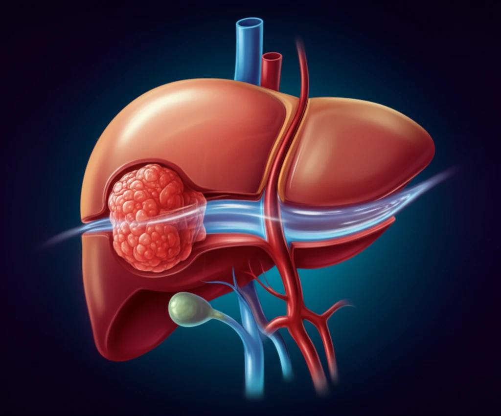
Liver cancer treatments have evolved significantly, and percutaneous imaging-guided ablation has become a common approach for treating hepatic malignancies. Ablation involves using heat to destroy tumor cells. Unfortunately, tumors near major blood vessels pose a challenge, because the blood flow dissipates the heat, reducing the effectiveness of the ablation. This phenomenon, known as the "heat-sink" effect, can lead to incomplete treatment and higher recurrence rates.
To combat the heat-sink effect, researchers have explored various strategies, including endovascular occlusion of the inferior vena cava (IVC) and alternative ablation techniques like microwave ablation (MWA) and irreversible electroporation (IRE). Hydrodissection, which involves injecting fluid between the tumor and adjacent structures, is now creating excitement.
A recent study highlights the use of retrohepatic hydrodissection to physically separate liver tumors from the IVC and hepatic veins. This innovative technique aims to minimize the heat-sink effect and improve ablation outcomes. Here’s how it works and what the initial results suggest.
How Does Retrohepatic Hydrodissection Work?
Retrohepatic hydrodissection involves injecting fluid into the retrohepatic space to create a physical barrier between the liver tumor and the inferior vena cava (IVC) and hepatic veins (HV). This separation is intended to reduce the heat-sink effect during thermal ablation, allowing for more effective tumor treatment and reducing the risk of vascular injury.
- Needle Placement: Two to three 22G spinal needles were inserted percutaneously under CT guidance to access the retrohepatic space.
- Fluid Injection: A mixture of 5% iodinated contrast and 0.9% saline solution was injected to dissect the space between the tumor and the IVC/HV. The mean volume of fluid used was 410 ml.
- Separation: The fluid injection physically separated the tumor from the IVC and the ostia of the right and middle hepatic veins. The average separation achieved was 9 mm.
- Ablation: After hydrodissection, microwave ablation (MWA) was performed to destroy the tumor cells.
What are the Potential Benefits and Future Directions?
Retrohepatic hydrodissection is a feasible technique to separate a tumour from the IVC and/or ostia of the HV, potentially limiting the heat-sink effect and reducing the risk of thrombosis. All lesions but one were completely ablated after one session at 3-month follow-up. The patient with residual tumour was successfully retreated. This advancement is an excellent step towards more effective and safer ablation of liver tumors located near critical blood vessels. More extensive studies are warranted to assess the efficacy on a long-term basis.