
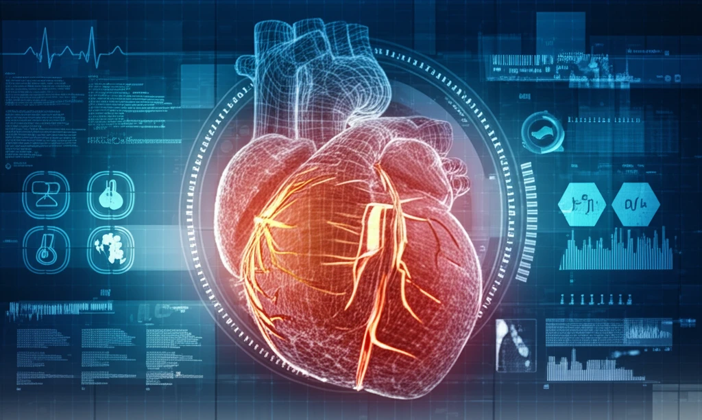
Heart Health Revolution: How 4D Cardiac Reconstruction is Changing Everything

"Discover the groundbreaking technology that's allowing doctors to see your heart like never before, leading to earlier diagnoses and more effective treatments."
Cardiovascular diseases remain a leading cause of mortality worldwide, making early detection and accurate diagnosis critical. Traditional methods, while effective to a degree, often fall short in capturing the intricate details of the heart's structure and function. But imagine if doctors could see your heart in motion, in high definition, revealing subtle anomalies that were previously undetectable. This is no longer a dream but a reality, thanks to advancements in 4D cardiac reconstruction.
Computed Tomography (CT) and Magnetic Resonance Imaging (MRI) have long been essential tools for visualizing the heart. However, these methods often provide static images, lacking the dynamic information needed to fully understand the heart's complex movements. Standard cardiac imaging provides valuable insights into wall thickness and overall function, detailed 3D models of cardiac structures have proven difficult to obtain due to data limitations, especially concerning finer details such as papillary muscles and trabeculae.
Now, with the advent of advanced multi-detector CT technologies, the possibility of capturing high-resolution, volumetric images of the heart in a single heartbeat is revolutionizing cardiac care. This breakthrough allows doctors to observe the heart's intricate movements and subtle changes throughout a cardiac cycle, which is invaluable for diagnosing and treating a range of cardiovascular conditions.
A Fast-Evolving Imaging Frontier
4D cardiac reconstruction is emerging as a key enabler of cardiac digital twins, yet researchers note that accurate 4D whole-heart mesh reconstruction from sparse cine MRI remains challenging due to limited 2D slice coverage and the complex coupling between cardiac shape and motion arxiv.org. To cope with data limitations, recent deep-learning frameworks reconstruct the 4D myocardium via decoupled motion and shape features, building an end-diastolic shape model to alleviate the problem of medical data scarcity openaccess.thecvf.com. Hardware advances have also contributed: 320 multi-detector CT now makes volumetric acquisition of high-resolution 4D cardiac images possible within a single heartbeat. The underlying physics is nonetheless demanding, since reconstructing a 3D image of the beating heart at a given phase requires a complete set of X-ray projection data representing that phase.
The Limits of ECG-Gated, Phase-Based Reconstruction
The standard method for 4D CT image reconstruction extracts individual cardiac motion phases and reconstructs them separately, but these reconstructions can suffer from undersampling artifacts because each phase relies on a limited number of projections academia.edu. Current 4D reconstruction methods typically rely on ECG-gating and assume periodic heart motion, an assumption that is often violated by arrhythmias or breathing, while C-arm CT adds further difficulty through long acquisition times and motion artifacts liner.com. A common procedural variant acquires volumetric data at different cardiac phase points using electrocardiogram-gated CT and alternates motion estimation with motion-compensated image reconstruction until convergence. These constraints are a major reason deep learning has entered the field, with reconstruction techniques now allowing unprecedented levels of data undersampling.
From Static CT to Beating-Heart Visualization
Foundational progress in this field has been driven largely by advances in CT hardware, with recent developments making the acquisition of high-resolution 4D cardiac images possible cs.rutgers.edu and 320 multi-detector CT systems enabling volumetric acquisition of such data within a single heartbeat academia.edu. Early frameworks built on these data reconstructed the 4D motion of the endocardial surface of the left ventricle across a full cardiac cycle. A further milestone was the introduction of a 4D cardiac reconstruction method that abandons the periodicity assumption, separating the estimation of the motion field from image reconstruction using an analytic motion compensation algorithm. Together, these developments mark the transition from static, single-phase imaging toward motion-aware, dynamic reconstruction of the heart.
The Power of 4D Cardiac Reconstruction
The core of this technological leap lies in the ability to reconstruct a comprehensive 4D motion model of the left ventricle (LV) from high-resolution CT images. This reconstruction framework captures the full 3D surfaces of complex anatomical features, including the often-elusive papillary muscles and ventricular trabeculae. For the first time, doctors can quantitatively investigate the functional significance of these structures in both healthy and diseased hearts.
- Enhanced Visualization: Captures details previously unseen, enabling a more comprehensive understanding of cardiac anatomy.
- Improved Diagnosis: Allows for earlier and more accurate detection of subtle abnormalities, leading to timely interventions.
- Personalized Treatment Planning: Provides detailed insights into individual heart function, facilitating tailored treatment strategies.
- Functional Insights: Enables the study of complex cardiac mechanics, potentially leading to new therapies for heart disease.
Active Research on ECG-Free Reconstruction
Recent research continues to push beyond the assumptions that constrain conventional 4D cardiac imaging. The field reports that 3D reconstruction of cardiac vasculature, such as the coronary arteries, using C-arm CT (rotational angiography) is an active and challenging field of research, with numerous publications exploring different reconstruction techniques. The sustained difficulty reflects the technical demands of imaging a moving organ with a rotating X-ray source, and the proliferation of published approaches suggests the community has not yet settled on a single standard solution.
When Periodic-Motion Assumptions Fail
A recurring criticism of conventional 4D cardiac reconstruction is its dependence on the assumption that heart motion is periodic. In the paper 'Towards 4-D Cardiac Reconstruction without ECG,' the authors introduce a method for cardiac 4D reconstruction that removes that periodicity assumption, proposing to separate estimation of the motion field from image reconstruction using an analytic motion compensation algorithm. The existence of such work underscores that periodicity-based reconstruction is viewed by researchers as a genuine limitation in real clinical settings, particularly where the heart rhythm is irregular. It also signals that reconstructing cardiac motion without an ECG signal remains an open and active challenge.
Phase-by-Phase vs. Whole-Cycle Reconstruction
Comparing 4D cardiac reconstruction methods highlights a fundamental design choice: whether to handle cardiac phases one by one or to solve for the entire heart cycle together. The 4D RecOnstructiOn using Spatial and TEmporal Regularization (4D ROOSTER) method exemplifies the latter, reconstructing all cardiac phases at once as a single 3D + time volume. By applying spatial and temporal regularization, it is designed to exploit information across the full cycle rather than treating phases independently, which points to meaningful differences from approaches that handle individual phases in isolation.
The Future of Cardiac Care is Here
4D cardiac reconstruction represents a paradigm shift in how we approach heart health. As the technology continues to evolve, we can expect even more detailed insights into cardiac function, leading to earlier diagnoses, more effective treatments, and ultimately, improved outcomes for patients with cardiovascular disease. With ongoing advancements, the goal is to capture more fine structures, such as valves and wall surfaces of all four chambers, providing an even more complete picture of the heart's intricate workings. This technology is not just about seeing the heart; it's about understanding it in a way we never thought possible.
Hardware and Software Converge
Taken together, progress in 4D cardiac reconstruction shows how advances in imaging hardware have converged with computational methods to move cardiac imaging from static snapshots toward dynamic, whole-beat visualization. Central to this progress is the development of 320 multi-detector CT technology, which has made the volumetric acquisition of high-resolution 4D cardiac images possible within a single heartbeat. Single-heartbeat acquisition matters because it reduces the motion-related misalignment that complicates multi-cycle, phase-based imaging. As the technique becomes increasingly feasible, high-resolution 4D cardiac imaging appears poised to become a more practical and routine clinical capability.
Robotics, Bioresorbables, and Personalization
Looking ahead, market analysis of the cardiac reconstruction devices space points to several defining trends: the rise of robotic-assisted surgeries, the use of bioresorbable materials, and an increasing focus on personalized medicine. These developments suggest that cardiac reconstruction is evolving not only as an imaging discipline but as part of a broader movement toward less invasive, patient-specific cardiovascular care. As these technologies mature, they are expected to shape how reconstruction devices are designed, manufactured, and adopted in clinical practice.
Systemic Barriers to Adoption
Beyond the technical details, widespread adoption of 4D cardiac reconstruction is likely to hinge on systemic factors such as the cost of advanced imaging hardware, the availability of specialized expertise, and integration into existing clinical workflows. Regulatory approval pathways and reimbursement structures will also influence how quickly these technologies reach routine care. As with many emerging imaging innovations, the gap between research demonstration and everyday clinical use remains substantial and will only be closed through coordinated effort across manufacturers, clinicians, and health systems.
What It Could Mean for Patients
Ultimately, the value of 4D cardiac reconstruction will be judged by its effect on patients and clinicians. If the technology matures as anticipated, it could give physicians a more complete view of how an individual heart moves and functions, potentially supporting earlier detection and more personalized treatment decisions. For patients, that could translate into better-informed procedures and more tailored follow-up care. These potential benefits, however, remain largely prospective, and realizing them will require careful validation in real-world clinical settings.