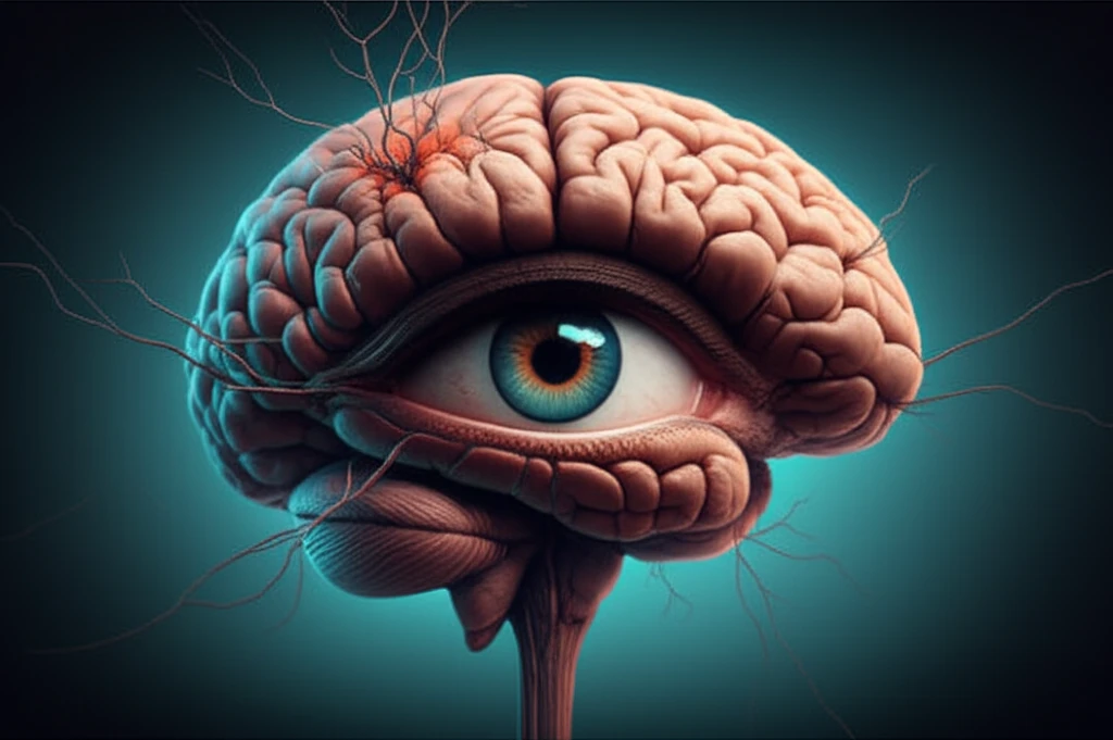
Glaucoma and the Brain: How One Eye's Trouble Affects Your Vision Center
"New research sheds light on the surprising stability of the brain's visual cortex, even when glaucoma affects one eye. Discover what this means for understanding and treating vision loss."
Glaucoma, a leading cause of blindness, is projected to affect over 80 million people worldwide by 2020. This condition damages the optic nerve, leading to vision loss that often starts with blind spots in the affected eye's field of view. While glaucoma's primary impact is on the eye, increasing evidence suggests that the effects of this neurodegenerative disease extend to the brain.
Specifically, research has shown that glaucoma can lead to decreased cell density and activity in the brain's visual processing centers, such as the lateral geniculate nucleus (LGN) and the primary visual cortex. These changes are thought to be due to the degeneration of nerve connections between the eye and the brain. However, it's been unclear whether this loss of input from the affected eye causes the visual cortex to functionally reorganize itself.
To investigate this, a recent study used functional magnetic resonance imaging (fMRI) to examine the brains of individuals with glaucoma in only one eye. The goal was to see if the visual cortex regions that had lost input from the glaucomatous eye showed an increased response to input from the unaffected eye. The results offer valuable insights into the brain's response to vision loss and the potential for therapies.
How Glaucoma Impacts the Visual Cortex: What the Study Revealed
The study compared nine participants with unilateral primary open-angle glaucoma (POAG) to a control group of four individuals without glaucoma. Participants with glaucoma had visual field defects in one eye, specifically in the central area of vision. Researchers used fMRI to measure brain activity while participants viewed visual stimuli with each eye separately.
- No Increased Response: The study found no evidence that the brain areas corresponding to the vision lost from the glaucomatous eye showed an increased response to the unaffected eye.
- Reduced Activation: Consistent with previous studies, there was a noticeable reduction in brain activity in both the primary (V1) and extrastriate (V2) visual cortex when participants viewed the stimuli through their glaucomatous eye. This confirms that fMRI can detect the impact of glaucoma on brain function.
- Stability in the Adult Brain: The results suggest a remarkable level of stability in the adult visual cortex, even in the face of nerve degeneration caused by glaucoma. This indicates that the adult brain may not reorganize itself as much as previously thought in response to this type of vision loss.
What This Means for Understanding Glaucoma and the Brain
This research suggests that in adults with unilateral glaucoma, the visual cortex maintains a surprising level of stability, without significant reorganization, as the primary and extrastriate visual cortex regions responding to the fellow eye were equivalent within the LPZ and the control ROIs.
The study indicates that the adult human visual cortex may not be as plastic as once believed in response to optic nerve neurodegeneration. Although there was a reduction in BOLD change in the LPZ ROIs for glaucomatous eye viewing, considerable residual activation was still evident for the high and medium contrast stimuli.
Further research should focus on examining the residual cortical responses near the LPZ to fully understand the origin of these signals. This is important for advancing new approaches, such as retinal prostheses, which may rely on the presence of functioning cortical cells even after extended visual deprivation.