Protected Heart: A visual representation of safeguarding cardiac health during cancer treatment.

Drug-Induced Cardiotoxicity: How to Protect Your Heart

"Protecting the Heart: New Strategies for Predicting and Preventing Cardiotoxic Side Effects of Chemotherapy"


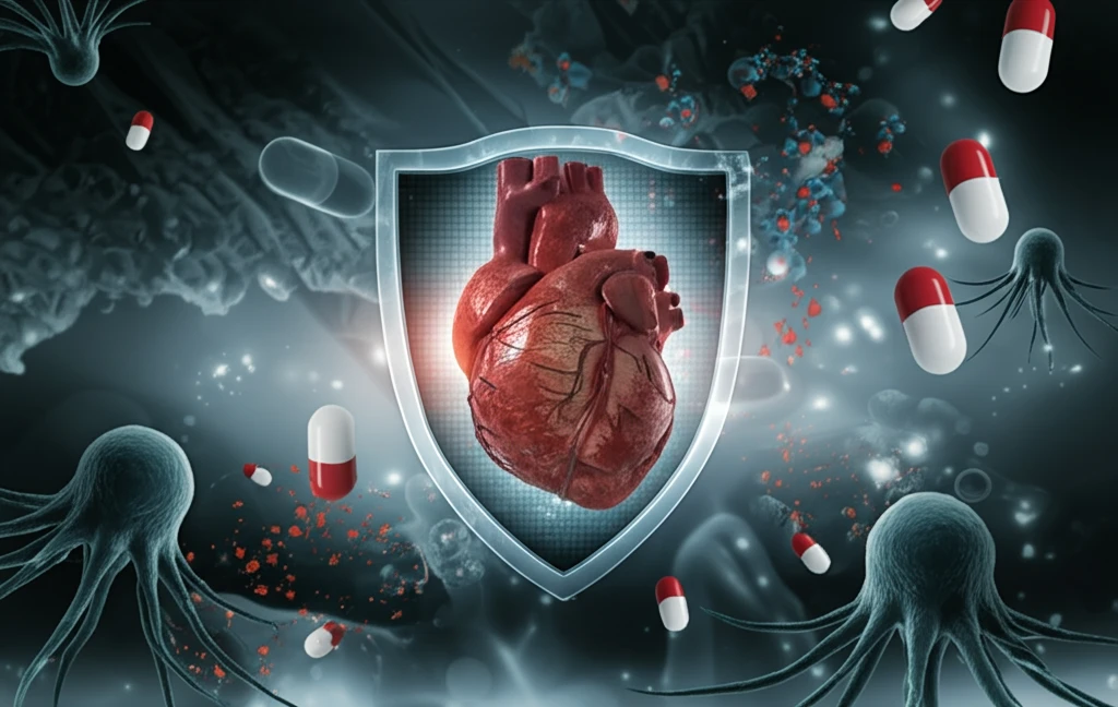
Cancer treatments, while life-saving, can sometimes have unintended consequences for the heart. Cardiotoxicity, or heart damage caused by drugs, is a significant concern in cancer therapy. Many chemotherapeutic agents can lead to a range of cardiovascular issues, from mild arrhythmias to severe heart failure. Understanding and mitigating these risks is crucial for improving patient outcomes and quality of life.

Researchers are actively investigating new methods to predict and prevent cardiotoxic side effects before they manifest in patients. These efforts include using advanced cell models and sophisticated computer simulations to assess the potential impact of drugs on the heart. By identifying risks early, doctors can make informed decisions about treatment plans, potentially adjusting dosages or selecting alternative therapies that are less harmful to the cardiovascular system.

This article delves into the latest research and strategies for protecting the heart during cancer treatment. We'll explore how scientists are using innovative approaches to identify cardiotoxic risks early, offering hope for safer and more effective cancer therapies.

Predicting Cardiotoxicity with hiPSC-Derived Cardiomyocytes

Protected Heart: A visual representation of safeguarding cardiac health during cancer treatment.

One promising approach involves the use of human induced pluripotent stem cell-derived cardiomyocytes (hiPSC-CMs). These cells, created from human stem cells, mimic the behavior of actual heart cells and can be used to study the effects of drugs in a controlled laboratory setting. Researchers can expose these cells to various chemotherapeutic agents and monitor their functional and structural responses to assess potential cardiotoxicity.

A recent study highlighted the potential of hiPSC-CMs to capture clinically relevant cardiotoxic effects of chemotherapeutics. The researchers treated hiPSC-CMs with drugs like lapatinib and sunitinib, which are known to have cardiotoxic potential. They then measured both functional toxicity (changes in cell activity) and structural toxicity (damage to cell structure) using sophisticated techniques like microelectrode array (MEA) impedance technology and cardiac Troponin I (cTnI) level measurements.

Here’s what the study revealed:
  • Functional Toxicity: Lapatinib altered the excitation-contraction coupling in cardiomyocytes, while sunitinib caused arrhythmic beating.
  • Structural Toxicity: Sunitinib induced a dose-dependent release of cTnI, a marker of heart damage, while lapatinib did not significantly affect cTnI levels.
  • Cell Viability: Sunitinib reduced cell viability, correlating with the increased release of cTnI.
These findings demonstrate that hiPSC-CMs can effectively model the diverse cardiotoxic effects of different drugs. By investigating both functional and structural toxicity, researchers gain a more comprehensive understanding of the potential risks associated with chemotherapeutic agents. This approach could lead to better risk assessment and more informed treatment decisions in the future.

Future Directions: Towards Safer Cancer Therapies

The development and application of advanced techniques like hiPSC-CMs and PBPK modeling represent significant strides in predicting and preventing drug-induced cardiotoxicity. As research progresses, these tools will likely become integral to the drug development process, helping to ensure that new cancer therapies are both effective and safe for the heart. By prioritizing cardiac safety, we can improve the overall well-being and long-term health of cancer patients.

About this Article -

This article was crafted using a human-AI hybrid and collaborative approach. AI assisted our team with initial drafting, research insights, identifying key questions, and image generation. Our human editors guided topic selection, defined the angle, structured the content, ensured factual accuracy and relevance, refined the tone, and conducted thorough editing to deliver helpful, high-quality information.See our About page for more information.

Everything You Need To Know

1

What exactly is cardiotoxicity in the context of cancer treatment, and why is understanding it so important?

Cardiotoxicity in the context of cancer treatment refers to the damage inflicted upon the heart as a consequence of drugs, especially chemotherapeutic agents. This damage can manifest as a range of cardiovascular issues, spanning from arrhythmias to heart failure. Understanding the mechanisms and risks associated with cardiotoxicity is crucial because it directly impacts patient outcomes and their overall quality of life during and after cancer therapy. Predicting and preventing cardiotoxicity allows for informed treatment decisions, potentially involving dosage adjustments or the selection of alternative therapies that are less harmful to the cardiovascular system. The long-term effects of cancer treatment may be improved, mitigating potential complications and improving overall well-being.

2

What are hiPSC-derived cardiomyocytes, and how are they used to predict cardiotoxicity in cancer treatments?

Human induced pluripotent stem cell-derived cardiomyocytes, or hiPSC-CMs, are cells created from human stem cells that mimic the behavior of actual heart cells. These cells are used in laboratory settings to study the effects of various drugs on heart tissue. Researchers expose hiPSC-CMs to chemotherapeutic agents and monitor their functional and structural responses, such as changes in cell activity and damage to cell structure, to assess the potential for cardiotoxicity. This approach enables a controlled and detailed examination of how specific drugs may harm the heart, providing insights that are difficult to obtain directly from patients. While the article doesn't mention other cell types used in cardiotoxicity studies, hiPSC-CMs are highlighted for their relevance to human cardiac tissue, offering a more predictive model compared to animal or generic cell lines.

3

According to the study, what specific effects did lapatinib and sunitinib have on hiPSC-CMs, and what do these effects indicate about cardiotoxicity?

The hiPSC-CM study revealed that lapatinib altered the excitation-contraction coupling in cardiomyocytes, indicating functional toxicity, while sunitinib caused arrhythmic beating. Sunitinib also induced a dose-dependent release of cardiac Troponin I (cTnI), a marker of heart damage, indicating structural toxicity, whereas lapatinib did not significantly affect cTnI levels. Additionally, sunitinib reduced cell viability, which correlated with the increased release of cTnI. These findings suggest that hiPSC-CMs can effectively model the diverse cardiotoxic effects of different drugs, and assessing both functional and structural toxicity provides a more comprehensive understanding of potential risks associated with chemotherapeutic agents. The study did not discuss the long-term effects of these drugs; further longitudinal studies would be needed to understand chronic impacts.

4

How do microelectrode array (MEA) impedance technology and cardiac Troponin I (cTnI) level measurements contribute to assessing cardiotoxicity using hiPSC-CMs?

Microelectrode array (MEA) impedance technology is used to measure functional toxicity, specifically changes in cell activity within hiPSC-CMs exposed to chemotherapeutic agents. It assesses how drugs affect the electrical properties and behavior of heart cells, providing insights into arrhythmic beating or altered excitation-contraction coupling. Cardiac Troponin I (cTnI) level measurements, on the other hand, quantify structural toxicity by detecting the release of cTnI, a marker of heart damage. Increased cTnI levels indicate damage to heart muscle cells. MEA impedance focuses on functional aspects, while cTnI measurements focus on structural damage. The techniques were useful for highlighting differing effects of lapatinib and sunitinib, as outlined in the study.

5

What are the future implications of using hiPSC-CMs and PBPK modeling in the development of safer cancer therapies, and what challenges might still exist?

The development and application of advanced techniques such as hiPSC-CMs and PBPK modeling represent significant advancements in predicting and preventing drug-induced cardiotoxicity. These tools are likely to become integral to the drug development process, helping to ensure that new cancer therapies are both effective and safe for the heart. By prioritizing cardiac safety, the overall well-being and long-term health of cancer patients can be improved. Future directions may involve refining these models, integrating them with other predictive methods, and conducting larger studies to validate their clinical utility. However, the article does not discuss regulatory pathways for adopting these technologies, which would be crucial for their widespread implementation.

Newsletter Subscribe

Subscribe to get the latest articles and insights directly in your inbox.