
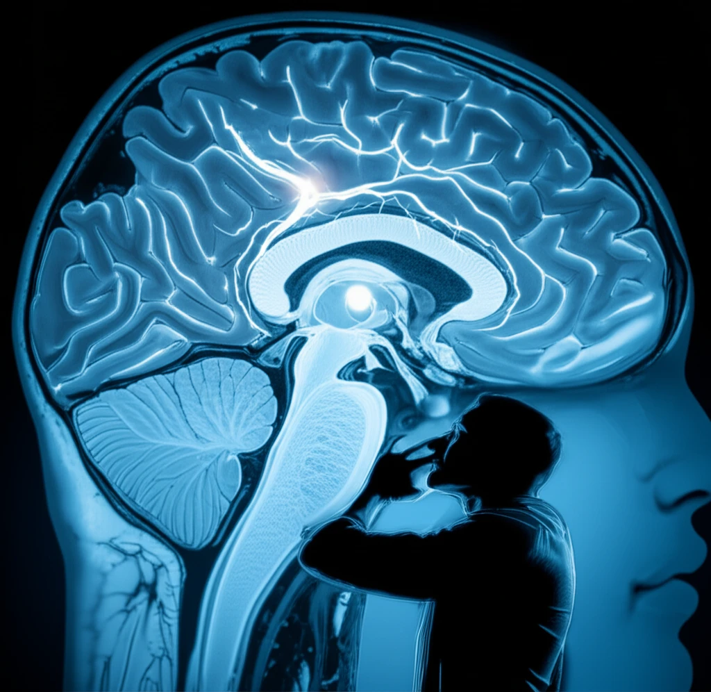
Deep Brain Stimulation Breakthrough: 7T MRI Pinpoints Key Target in Parkinson's

"High-resolution imaging combined with histology offers a new level of precision for targeting the pedunculopontine nucleus in DBS."
Deep brain stimulation (DBS) has emerged as a promising treatment for Parkinson's disease, particularly for symptoms that don't respond well to medication. One key target for DBS is the pedunculopontine nucleus (PPN), a region in the brainstem involved in motor control, balance, and gait. However, accurately locating the PPN for DBS can be challenging due to its small size and the limitations of conventional MRI techniques.
Traditional MRI scans often struggle to clearly distinguish the PPN from surrounding brain structures, making precise targeting difficult. Recent research has focused on using advanced imaging techniques, such as diffusion tensor imaging (DTI), to visualize the white matter tracts that surround the PPN and use them as landmarks. DTI measures the direction and magnitude of water diffusion in the brain, providing detailed information about the organization of white matter.
A groundbreaking study published in Surgical and Radiologic Anatomy has taken this approach a step further by combining high-resolution 7T MRI with histological validation. The researchers were able to precisely identify the PPN and its surrounding white matter tracts in a post-mortem brain, paving the way for more accurate and effective DBS targeting in Parkinson's disease.
DBS Reach and Parkinson’s Burden
Deep brain stimulation (DBS) is an approved treatment option in the United States for Parkinson’s disease, essential tremor, dystonia, obsessive-compulsive disorder and epilepsy. A review estimates that Parkinson’s disease affects 1 million people in the United States and 10 million worldwide, and links the condition to the loss or death of dopaminergic neurons in the substantia nigra. Recent research has also examined whether racial and socioeconomic disparities in DBS use have improved over time, while a 2025 cohort study evaluated five-year outcomes and safety for subthalamic nucleus DBS.
Established Treatment, Ongoing Limits
DBS is generally understood as a surgical treatment used when Parkinson’s symptoms are not adequately controlled with medication. The approach requires careful clinical decision-making and has limitations related to surgery, targeting and subsequent therapy adjustment. Because no source material was provided for this subsection, these points should be treated as a general overview rather than a detailed comparison of techniques or outcomes.
From Experiment to Established Therapy
Historical reviews describe DBS as the result of contributions from neurosurgeons, neurologists and neurophysiologists across the 20th and early 21st centuries. One account emphasizes the plurality of interests involved in developing and stabilizing DBS technology, while another traces the field’s origin and evolution. Over the last two decades, DBS has produced positive results for medically refractory Parkinson’s disease, essential tremor and dystonia, and the last 50 years have brought rapid improvements in its safety and efficacy.
Unlocking the PPN: How 7T MRI and DTI Improve Targeting
The study, led by researchers at Radboud University Medical Center and Maastricht University Medical Center, used a 7T MRI scanner – a powerful imaging tool that provides exceptionally detailed images – to scan a post-mortem brain. They also used DTI to map the white matter tracts surrounding the PPN.
- DTI and White Matter Tracts: DTI allowed them to visualize the superior cerebellar peduncle (SCP) and the medial lemniscus (ML), two major white matter tracts that border the PPN.
- FA Maps: They used fractional anisotropy (FA) maps, which are derived from DTI data and show the degree to which water diffusion is restricted in a particular direction. The PPN appeared as an area of low FA, surrounded by the high FA values of the white matter tracts.
- Histological Validation: The histological sections confirmed the location of the PPN as a distinct area between the SCP and the ML.
A Field Still Advancing
Recent reviews suggest that DBS research continues to examine targeting, stimulation strategies and treatment optimization. The direction of this work appears to include more individualized approaches and improved use of anatomical, functional and imaging information. Because no source material was provided for this subsection, specific claims about the latest studies or measured breakthroughs cannot be established here.
Benefits Must Be Weighed Against Risks
A 2024 systematic review identified seven trials involving 1,125 participants randomized to DBS with antiparkinsonian drugs versus antiparkinsonian drugs alone, specifically assessing benefits and harms. DBS is established mainly for motor symptoms resistant to pharmacological therapy, but reviews continue to examine how targeting and imaging might improve results. Adaptive DBS is described as promising, yet its limitations still require further research and development, so it should not be treated as a fully resolved solution.
No Direct Comparison Provided
DBS can be compared with medication-based care in broad clinical discussions, particularly when symptoms remain resistant to pharmacological treatment. However, the supplied material contains no source specifically designed to compare DBS with another treatment across effectiveness, safety or quality-of-life measures. Any stronger comparative conclusion would therefore require additional evidence.
The Future of DBS: Personalized Targeting for Better Outcomes
This study provides strong evidence that 7T MRI, combined with DTI, can be used to precisely identify the PPN and its surrounding white matter tracts. This has significant implications for deep brain stimulation in Parkinson's disease, as it could lead to more accurate targeting and improved clinical outcomes.
Precision and Personalization
Taken together, the available material presents DBS as an established intervention whose development has depended on technical innovation and multidisciplinary expertise. It also indicates that improving anatomical, functional and imaging approaches may help optimize treatment. Without dedicated source material for this subsection, broader expert conclusions about the specific 7T MRI finding should remain cautious.
Toward Adaptive and Better-Mapped Stimulation
Future DBS research includes brain mapping for more precise targeting and adaptive DBS that listens to brain signals and responds like a pacemaker. A 2026 Clinical Outlook addresses adaptive DBS principles, current clinical recommendations, practical implementation and remaining challenges. These developments point toward more personalized therapy, but the supplied sources also emphasize that important implementation and treatment limitations remain.
Questions of Access and Implementation
The introduction of adaptive DBS raises broader questions about who should receive it, why it should be used and how treatment should be delivered. Reviews of DBS also focus on advanced anatomical, functional and imaging approaches for patients whose motor symptoms remain resistant to pharmacological therapy. These issues show that progress involves not only technical capability but also decisions about eligibility, implementation and equitable access.
Access Beyond the Operating Room
A 2026 multicenter assessment reports that remote, internet-based DBS programming for Parkinson’s disease can accelerate postoperative clinical benefits by improving access to therapy adjustments. A separate patient account describes bilateral subthalamic nucleus DBS surgery in May 2020 using Vercise Gevia with Cartesia directional leads from Boston Scientific, followed by activation the next day and substantially reduced medication before surgery. These examples illustrate that real-world impact depends not only on implantation but also on continuing programming, access and patient experience.
By using these advanced imaging techniques, neurosurgeons may be able to personalize DBS therapy by tailoring the placement of electrodes to the individual patient's anatomy. This could result in greater symptom relief and fewer side effects.
While this study was conducted on a post-mortem brain, the researchers emphasize the need for in vivo validation to confirm these findings in living patients. Future research will focus on translating this technology to the clinic and evaluating its effectiveness in improving DBS outcomes for people with Parkinson's disease. The future of DBS lies in precision, and this research brings that future closer to reality.