
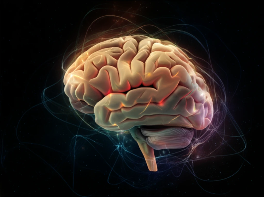
Decoding Your Brain: How Scientists Are Mapping the Amazing Cerebellum

"From movement to memory, discover how cutting-edge research is unlocking the secrets of this essential brain region."
Imagine a tiny, walnut-shaped structure tucked away at the back of your head. This unassuming part of your brain, the cerebellum, is a powerhouse of activity, orchestrating everything from your graceful dance moves to your ability to learn a new language. For years, scientists have been fascinated by the cerebellum, and now, with the help of advanced imaging techniques, they're unraveling its intricate workings in unprecedented detail.
This article will take you on a journey through the latest research, offering a clear and engaging look at how scientists are mapping the cerebellum. We'll explore how these maps are created, what they reveal about the brain's functions, and why this knowledge is so important for understanding both healthy brain function and a range of neurological conditions.
This is not a highly technical journal, but an exploration of science. We are going to simplify the concepts. You will be able to understand what the research means, and why it matters to you.
A Repeating Circuit Beyond Motor Control
The cerebellar cortex has a complex but remarkably homogeneous cellular organization repeated across its extent. Its connections with the cerebral cortex are not limited to the long-recognized motor and premotor areas; different cerebellar regions have specific feedforward and feedback connections. A review describes the cerebellar circuit as mostly feedforward from mossy fiber inputs toward deep nuclear outputs, with expansion to granule cells and compression toward output cells.
From Hidden Structure to Broader Functions
The cerebellum has long been recognized as a distinct brain subdivision, though in humans it is hidden from a top view by the occipital lobe. The species difference in whether the cerebral cortex covers the cerebellum was noted in the historical account as part of the background to Vesalius’s observations. More recent support for broader cerebellar roles comes from recognition of cerebellar cognitive affective syndrome, connectional neuroanatomy, and anatomical, task-based, and functional-connectivity MRI. A 2025 study mapped cerebellar topography with fine temporal granularity and reported hierarchical cerebellocortical functional connectivity from infancy.
The Cerebellum's Hidden Power: What Does It Do?
The cerebellum, often called the "little brain," is responsible for a wide array of functions that go far beyond just movement. It acts as a central hub for coordinating our actions, thoughts, and emotions. It receives information from various parts of the brain and spinal cord, processes it, and then sends signals back to these areas to fine-tune our responses.
- Motor Control: The cerebellum is the master conductor of movement, ensuring smooth, coordinated actions. It helps us walk, write, and perform any skill that requires physical coordination.
- Cognitive Functions: Recent research has revealed that the cerebellum plays a vital role in learning, memory, attention, and even language processing.
- Emotional Regulation: Studies suggest that the cerebellum is involved in processing and regulating emotions, contributing to our overall emotional well-being.
Genetics, Symptoms, and Mapping Tools
A twin study of cerebellar gray matter volume reported that additive genetic factors explained about 45% of voxel-level variance, with minimal shared environmental effects; voxel-wise mapping also found within-lobule heterogeneity not captured by averaging across lobules. A secondary analysis from the INSPiRE-TMS trial associated broadly defined post-stroke dizziness and vertigo with a lesion network involving cerebellar and occipital regions. Separately, the SUIT toolbox offers a surface-based cerebellum representation for displaying volume-averaged data on a group flatmap.
Interpretation Depends on Methods
A paper on surface-based display of volume-averaged cerebellar imaging data cautions that visualization techniques can strongly influence how data are understood, so the technique’s limitations matter. Research on cerebellar functional gradients identifies a further open question: whether inter-subject variability in those gradients can predict task performance, diagnosis, prognosis, or treatment response. The cited material presents these as limitations and future research questions, not established predictive outcomes.
Contrasting Measures and Functional Atlases
A report on magnesium-deprived mice found significantly smaller magnesium values in the cerebellum than in the brain. In a separate line of work, a 2024 study developed a probabilistic human cerebellum atlas by combining multiple task-based datasets to map its mosaic of functional regions. The atlas authors chose task-based data alone for the final atlas, with the stated goal of comprehensively mapping motor and non-motor cerebellar regions.
A Brighter Future for Brain Health
As scientists continue to map the cerebellum, we can expect even more breakthroughs in our understanding of the brain. This research not only enhances our knowledge of healthy brain function but also paves the way for new treatments for neurological conditions. By exploring the power of the "little brain," we're taking significant strides towards a healthier and more informed future.
A Growing Imaging Toolkit
A 2023 review reports that many image-processing tools needed for detailed analysis of cerebellar structure and function are available. It describes capabilities ranging from denoising and super-resolution to cortical mapping. The review’s summary points to a broad toolkit for cerebellar imaging analysis, without specifying a single method as sufficient for every research question.
Sharper Views of Cerebellar Networks
A paper on advancing cerebro-cerebellar network imaging with ultra-high-field methods suggests that future developments may provide access to cerebellar substructures distinguished by myelination differences. The authors anticipate that this could offer further insight into cerebro-cerebellar networks. Separately, a 2024 human cerebellum atlas paper notes that the cerebellum is activated by a wide variety of cognitive and motor tasks and that previous functional atlases relied on single task-based or resting-state fMRI datasets.
Separating Anatomy from Confounds
A 2025 study used voxel-based lesion-symptom mapping in children and young adults who had undergone cerebellar tumor resection. The approach was used to control for covariates and evaluate causal relationships between brain anatomy and behavioral performance. The stated aim was to help disentangle the anatomical substrates of motor and cognitive functions.