
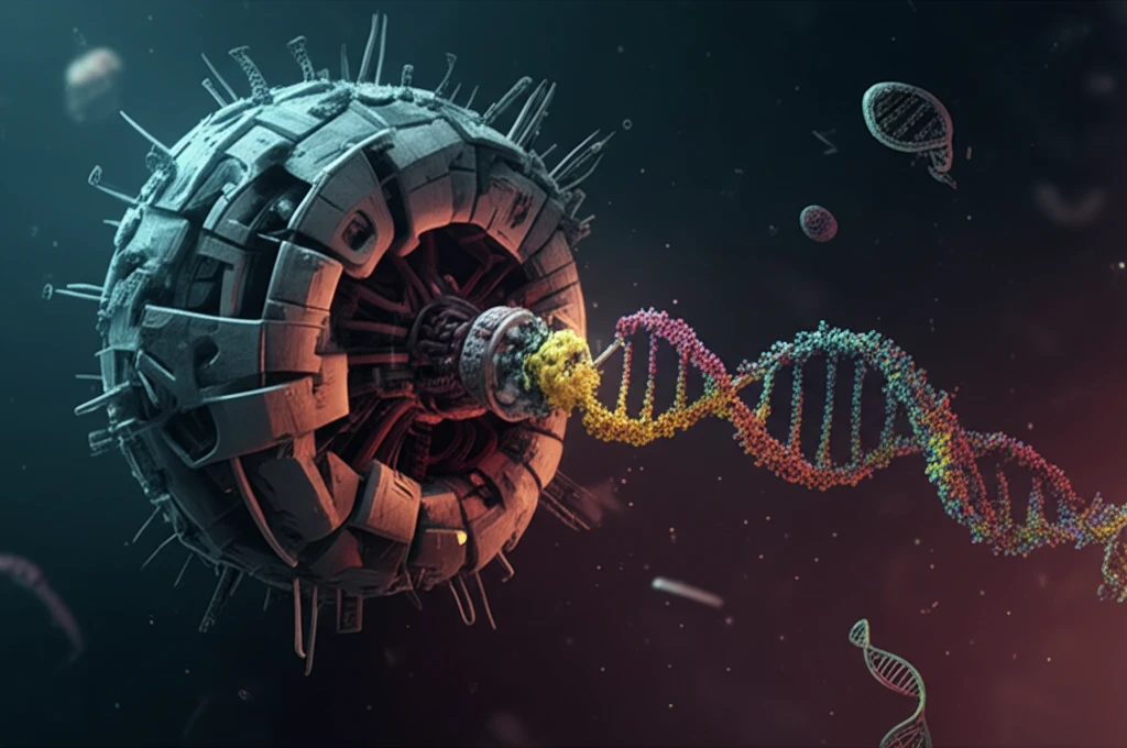
Decoding Viral RNA: How Ribosomes Remodel Genetic Messages

"Unlocking the Secrets of RNA Structure to Understand Viral Gene Regulation"
Gene expression, the process by which our cells read and use the instructions encoded in our DNA, is a tightly controlled affair. At the heart of this process lies RNA, a versatile molecule that acts as an intermediary between DNA and protein synthesis. RNA isn't just a passive carrier of information; it's an active player, capable of folding into intricate three-dimensional structures that dictate its function.
Structured RNA elements, programmed RNA conformational changes, and interactions between different RNA domains underlie many modes of regulating gene expression, mandating studies to understand the foundational principles that govern these phenomena. Viruses, masters of efficiency, often exploit these structural intricacies to hijack the host cell's machinery for their own replication.
Now, a team of scientists has delved into the complex world of viral RNA, uncovering a fascinating mechanism by which ribosomes, the protein-synthesizing workhorses of the cell, can physically reshape viral RNA structure. This discovery, focused on the turnip yellow mosaic virus (TYMV), sheds light on how viruses fine-tune gene expression and opens up new avenues for antiviral drug development.
Current Statistics & Impact
High-resolution structural data from cryo-electron microscopy have revealed the intricate static and dynamic nature of RNA structures in ribosomes, with the human 80S ribosome structure resolved to 3.6 Å average resolution and 2.9 Å in stable regions. The Ribosome Atlas now provides an interactive resource for exploring ribosomal RNA diversity across microbial life, integrating large-scale phylogenies and structural visualizations. These advances enable detailed analysis of rRNA evolution and ribosomal variation across taxonomic levels, highlighting the complexity of RNA structures as they perform their functions in translation.
Standard Approach, Accepted Methods & Their Limitations
Traditional approaches to studying ribosomal RNA structure have relied on comparative sequence analysis, thermodynamic folding algorithms, and biochemical probing methods, each with inherent limitations in capturing dynamic conformational changes. Comparative analysis depends on covariation signals that may be weak or absent in rapidly evolving regions, while thermodynamic models often fail to account for noncanonical interactions and long-range tertiary contacts. Experimental methods such as chemical probing provide valuable constraints but can be limited by resolution and the challenge of capturing transient structural states in living cells.
Historical Perspective, Milestones, Foundational Discoveries
The ribosome evolved before the last universal common ancestor, with evidence from primary sequences, high-resolution structural studies, and functional properties providing insights into this evolutionary history linked to the origins of the genetic code and chirality. George Emil Palade first observed ribosomes in 1955, launching decades of structural and functional research into ribosome biogenesis involving rRNA synthesis, processing, and ribosomal protein assembly. The peptidyl transferase center (PTC) represents the ancient catalytic core where peptide bonds are formed, and its origin and early evolution can be elucidated through analysis of ribosomal proteins' taxonomic block structures and their RNA interactions.
How Do Ribosomes Change RNA Structure?
The study focuses on the 3'-untranslated region (UTR) of TYMV RNA, a region that doesn't code for proteins but plays a crucial role in regulating translation, the process of protein synthesis. The researchers discovered that the 3'-UTR acts as a sensor, responding to the presence of ribosomes by undergoing conformational changes.
- Ribosome-Induced Changes: The arrival of a ribosome near the UPD causes it to unfold.
- Communication Conduit: This unfolding propagates through a "spine" of continuously stacked bases to the TLS.
- Translation Adjustment: The TLS, in turn, adjusts translation efficiency based on the status of the UPD.
Latest Research and Reviews
Recent reviews highlight emerging methodologies for transcriptome-scale determination of RNA structures in living cells, encompassing both mRNAs and noncoding RNAs. Deep learning approaches are beginning to address RNA structure prediction challenges, though limited training data, complex noncanonical interactions, and conformational flexibility remain significant hurdles. Innovative techniques are uncovering distinct ribosome subtypes with significant structural variations and expression patterns, revealing how ribosomal RNA diversity impacts ribosome structure and its implications for health and disease.
Counter Arguments and Failures
Despite advances in structural biology, significant gaps remain in understanding how dynamic RNA remodeling occurs during viral infection and ribosome hijacking. Comparative structure models based on covariation analysis can produce inaccurate predictions when evolutionary signals are weak or when convergent evolution obscures true structural relationships. Thermodynamic folding algorithms often fail to predict biologically relevant structures due to their inability to fully capture kinetic folding pathways, protein cofactor interactions, and the influence of the cellular environment on RNA conformation.
Comparative Analysis
Comparative analysis of ribosomal RNA structures relies on the principle that covariation in base-paired positions across homologous sequences reveals conserved secondary structures, offering an alternative to thermodynamic folding algorithms. The Comparative RNA Web (CRW2) provides covariation-based secondary structures for rRNAs and group I and II introns, enabling large-scale comparative sequence and structure analysis. Recent reviews emphasize the integration of thermodynamic and comparative approaches for RNA-RNA interaction prediction, highlighting how these complementary methods improve accuracy in predicting RNA secondary structures and intermolecular interactions.
Why Is This Important?
This research provides a crucial understanding of how viruses manipulate RNA structure to control gene expression. By uncovering the interplay between ribosomes and viral RNA, scientists can now explore new strategies for disrupting viral replication. Imagine developing antiviral drugs that target the flexible linker between the UPD and TLS, preventing the ribosome from triggering the necessary structural changes. Or perhaps, designing molecules that stabilize the UPD, keeping the TLS in an 'off' state, effectively silencing viral protein production. The possibilities are vast, and this study serves as a critical stepping stone towards innovative antiviral therapies.
Synthesis & Expert Commentary
Deep learning has revolutionized protein structure prediction, but RNA presents unique challenges including limited training data, complex noncanonical interactions, and conformational flexibility that hinder direct application of similar approaches. High-throughput sequencing-based methods now enable transcriptome-scale determination of RNA structures in living cells, providing dynamic views of the RNA structurome for both mRNAs and noncoding RNAs. These advances collectively suggest that integrating experimental probing data with computational modeling will be essential for decoding how ribosomes remodel viral RNA messages during infection.
Future Outlook & Next Frontiers
Future research will likely focus on integrating time-resolved structural methods with single-molecule approaches to capture the dynamic remodeling of viral RNA by ribosomes in real time. Advances in cryo-EM and computational modeling may enable visualization of transient ribosomal intermediates during viral RNA translation and frameshifting events. Emerging technologies for in vivo RNA structure probing combined with ribosome profiling could reveal how specific RNA structural elements direct ribosomal behavior during viral infection.
Broader Context & Systemic Challenges
Recent work reveals how ribosomal RNA diversity impacts ribosome structure and function, with innovative methodologies uncovering distinct ribosome subtypes exhibiting significant structural variations and expression patterns relevant to health and disease. The emergence of ribosome structure and its interactions with substrates such as tRNAs represents a central process in cellular biology that viruses exploit for their own replication. Understanding ribosomal heterogeneity and its functional consequences presents both a fundamental biological question and a potential therapeutic target for antiviral strategies.
The Human Element & Real-World Impact
Understanding how ribosomes decode and remodel viral RNA has direct implications for developing antiviral therapies that target virus-specific translation mechanisms without disrupting essential host protein synthesis. Ribosomal heterogeneity and specialized ribosome subtypes may explain tissue-specific viral tropism and differential disease outcomes among individuals. Insights into viral RNA-ribosome interactions could inform the design of mRNA therapeutics and vaccines that optimize translation efficiency while minimizing unintended immune activation.