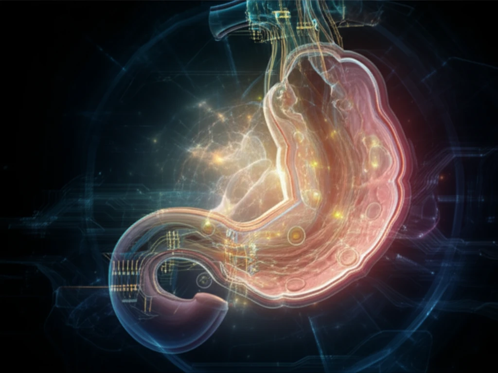
AI-powered oesophagus cancer imaging

Decoding Oesophageal Cancer: Can AI-Powered Imaging Predict Your Outcome?

"A new study explores how radiomics, using AI to analyze PET scans, could revolutionize oesophageal cancer treatment by improving risk prediction and personalizing patient care."


Oesophageal cancer, a formidable adversary with a survival rate of approximately 15% over five years, demands innovative approaches to improve patient outcomes. Current treatment strategies rely heavily on radiological staging to determine the prognosis and guide management plans. However, the limited progress in survival rates over recent decades underscores the need for enhanced methods that can more accurately predict how individual patients will respond to treatment.

The integration of artificial intelligence (AI) into medical imaging, known as radiomics, offers a promising avenue for advancing cancer care. Radiomics involves the automated extraction of quantitative features from radiological images, such as PET scans, to create detailed profiles of tumors. These profiles can potentially reveal hidden patterns and predict treatment responses that are not discernible through traditional visual assessment. By leveraging AI to analyze vast amounts of imaging data, researchers aim to develop more precise prognostic models that can personalize treatment strategies and improve patient outcomes.

A recent study has explored the potential of radiomics in oesophageal cancer by attempting to validate a previously developed prognostic model that incorporates quantitative PET image features. While the initial model did not demonstrate significant predictive power in the external validation cohort, the study shed light on the challenges of translating AI-driven models across different medical centers and imaging protocols. This exploration highlights the ongoing efforts to refine and standardize radiomic approaches, paving the way for more effective and personalized cancer treatments.

What is Radiomics and How Can It Help Oesophageal Cancer Patients?

AI-powered oesophagus cancer imaging

Radiomics is a cutting-edge field that combines the power of artificial intelligence with medical imaging to extract a wealth of information from radiological scans. By using sophisticated algorithms, radiomics can identify and quantify a variety of features within tumors, including their size, shape, texture, and metabolic activity. These features, often invisible to the naked eye, can provide valuable insights into the tumor's characteristics and behavior.

In the context of oesophageal cancer, radiomics offers the potential to:

  • Improve Risk Stratification: Better predict which patients are at higher risk of recurrence or treatment failure.
  • Personalize Treatment Decisions: Tailor treatment plans based on individual tumor characteristics.
  • Enhance Staging Accuracy: Provide more precise information about the extent and spread of the cancer.
  • Monitor Treatment Response: Track changes in the tumor over time to assess how well the treatment is working.
By integrating radiomic data with clinical information, doctors can gain a more comprehensive understanding of each patient's unique situation. This holistic approach can lead to more informed decisions about treatment options, ultimately improving the chances of successful outcomes and enhanced quality of life.

The Future of Radiomics in Cancer Care

While challenges remain in standardizing radiomic approaches and ensuring their generalizability across different medical centers, the potential benefits of AI-driven imaging in cancer care are undeniable. As technology advances and data collection becomes more streamlined, radiomics is poised to play an increasingly important role in personalizing treatment strategies and improving outcomes for patients with oesophageal cancer and other malignancies. By continuing to refine and validate these innovative techniques, we can move closer to a future where cancer treatment is tailored to the individual, maximizing the chances of success and minimizing the burden of this devastating disease.

About this Article -

This article was crafted using a human-AI hybrid and collaborative approach. AI assisted our team with initial drafting, research insights, identifying key questions, and image generation. Our human editors guided topic selection, defined the angle, structured the content, ensured factual accuracy and relevance, refined the tone, and conducted thorough editing to deliver helpful, high-quality information.See our About page for more information.

This article is based on research published under:

DOI-LINK: 10.1016/j.radonc.2018.10.033, Alternate LINK

Title: External Validation Of A Prognostic Model Incorporating Quantitative Pet Image Features In Oesophageal Cancer

Subject: Radiology, Nuclear Medicine and imaging

Journal: Radiotherapy and Oncology

Publisher: Elsevier BV

Authors: Kieran G. Foley, Zhenwei Shi, Philip Whybra, Petros Kalendralis, Ruben Larue, Maaike Berbee, Meindert N. Sosef, Craig Parkinson, John Staffurth, Tom D.L. Crosby, Stuart Ashley Roberts, Andre Dekker, Leonard Wee, Emiliano Spezi

Published: 2019-04-01

Everything You Need To Know

1

What is radiomics, and how does it aim to improve oesophageal cancer treatment?

Radiomics is a field that leverages artificial intelligence (AI) to analyze medical images, such as PET scans, extracting quantitative features from tumors. In oesophageal cancer, radiomics aims to improve risk prediction, personalize treatment decisions, enhance staging accuracy, and monitor treatment response. By creating detailed profiles of tumors, radiomics can reveal hidden patterns and predict treatment responses that are not discernible through traditional visual assessment, ultimately aiming to improve patient outcomes.

2

How does radiomics use PET scans in the context of oesophageal cancer?

Radiomics utilizes PET scans by employing AI to extract a wealth of information from these radiological images. Sophisticated algorithms analyze the scans to identify and quantify various features within the oesophageal tumors, including size, shape, texture, and metabolic activity. These features provide valuable insights into the tumor's characteristics and behavior, which can be used for improved risk stratification, personalized treatment, and monitoring of treatment responses.

3

What are the main challenges in applying radiomics to oesophageal cancer treatment?

One of the main challenges in applying radiomics to oesophageal cancer treatment is standardizing radiomic approaches and ensuring their generalizability across different medical centers and imaging protocols. A recent study highlighted the difficulty of translating AI-driven models across different settings. These challenges involve refining and validating radiomic techniques to achieve consistent and reliable results that can be widely applied for effective and personalized cancer treatments.

4

How can radiomics lead to more personalized treatment strategies for oesophageal cancer?

Radiomics can lead to more personalized treatment strategies by integrating radiomic data with clinical information to gain a more comprehensive understanding of each patient's unique situation. This holistic approach allows doctors to tailor treatment plans based on individual tumor characteristics, improving risk stratification, enhancing staging accuracy, and monitoring treatment response. By using AI to analyze data from PET scans, radiomics can help customize treatment options and improve the chances of successful outcomes, ultimately enhancing the quality of life.

5

Besides oesophageal cancer, what other types of cancer can radiomics potentially help treat?

While the study focuses on oesophageal cancer, the potential benefits of AI-driven imaging, like radiomics, extend to other malignancies as well. Radiomics can be applied to various cancers where medical imaging, such as PET scans, is used for diagnosis, staging, and treatment monitoring. As the technology advances and data collection becomes more streamlined, radiomics is poised to play an increasingly important role in personalizing treatment strategies and improving outcomes for patients with different types of cancer.

Newsletter Subscribe

Subscribe to get the latest articles and insights directly in your inbox.