
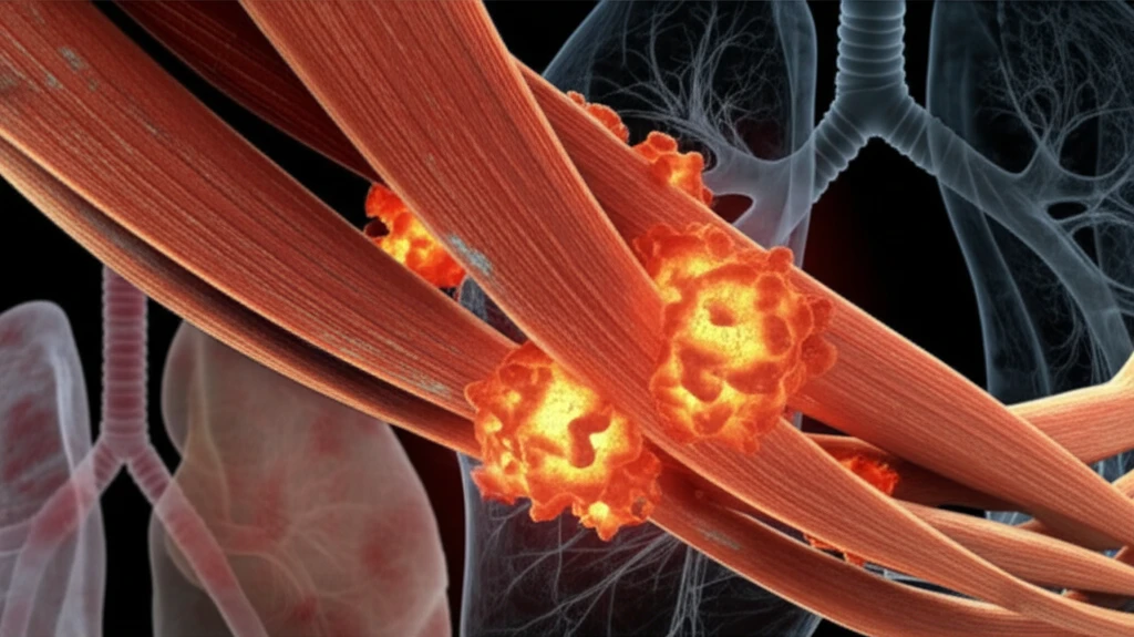
Decoding Myositis: A Practical Guide to Spotting and Managing Muscle Inflammation

"Early suspicion and comprehensive management are key to navigating the complexities of myositis, a group of inflammatory muscle diseases."
Myositis refers to a group of inflammatory conditions affecting the skeletal muscles. While all myositis share muscle inflammation as a core feature, the severity and impact can vary greatly, ranging from subtle, asymptomatic cases to severe, debilitating muscle weakness. Adding to the complexity, myositis often presents as part of a systemic illness, affecting multiple organs beyond just the muscles.
This widespread impact makes early detection and accurate diagnosis critical. Recognizing the signs, understanding the diagnostic pathways, and implementing appropriate management strategies are key to improving outcomes for individuals with myositis. The good news is that advances in identifying specific auto-antibodies have significantly improved diagnosis and classification, though muscle biopsy remains essential in many cases.
This article provides a practical guide to understanding myositis, drawing on the latest research to explain how to suspect, confirm, and manage the condition. We'll explore the key signs and symptoms, diagnostic tools, and management strategies to empower you with the knowledge you need.
How Common Is Myositis?
Idiopathic inflammatory myopathies (IIM) are described as rare autoimmune diseases, with reported incidence rates ranging from 0.2 to 2 cases per 100,000 person-years. A separate estimate from Hospital for Special Surgery puts the number of people living with myositis in the United States at about 50,000, while noting that the exact number is unknown. These figures describe different measures—incidence and estimated prevalence—rather than directly conflicting counts.
Biopsy and Imaging in Evaluation
Muscle biopsy is used in evaluating myositis and its differential diagnoses in adults and children, and available techniques include open surgical biopsy, needle muscle biopsy, and conchotome forceps procedures, each with distinct advantages and disadvantages. For acute-onset myositis, MRI and ultrasound can detect muscle inflammation and edema. In chronic myositis such as inclusion body myositis (IBM), ultrasound may help assess proximal and distal muscles together and evaluate asymmetry.
From Muscle Inflammation to Myopathy
Myositis refers to a myopathy in which muscle inflammation is a prominent feature; when its cause is unknown, it is classified as an idiopathic inflammatory myopathy. A separate overview defines myositis as inflammation of a muscle, especially a voluntary muscle, and describes pain, tenderness, swelling, or weakness as possible features. Historical accounts note that myositis-like conditions were recognized across centuries, although ancient and medieval physicians lacked a framework to distinguish them from other causes of muscle pain and weakness.
When to Suspect Myositis: Recognizing the Clues
Because myositis can manifest in diverse ways, vigilance is key. Suspect myositis when encountering certain clinical scenarios, especially those involving muscle-related symptoms, skin manifestations, lung involvement, or joint issues. Let's break down specific signs that should raise suspicion:
- Difficulty rising from a seated position (sign of the stool).
- Using hands to push oneself up from the floor (Gowers' sign).
- Inability to hold limbs elevated against gravity (Barré sign).
Refining Myositis Subtypes
A retrospective, multicentre study focuses on immune-mediated necrotizing myopathy (IMNM), described as a recently recognized inflammatory muscle disorder. Its characteristic features include subacute proximal muscle weakness, elevated serum creatine kinase (CK), and muscle-fiber necrosis with relatively little inflammation on histopathology. A 2025 comprehensive review reports that histopathological and immunochemical analysis of inflammatory muscle infiltrates, alongside discovery of new auto-antibodies, has helped identify more homogeneous myositis subsets.
Limits in Classification and Assessment
An England population-based cohort study covering 2002–2021 notes that its “other IIM” group remains heterogeneous and may include polymyositis, unspecified myositis, overlap myositis, antisynthetase syndrome, and IMNM. For unspecified myositis, the study additionally required coded evidence of interstitial lung disease (ILD), a limitation of its case definition. Separately, a review of MRI and muscle imaging notes that assessing disease activity and muscle damage is difficult because strength evaluations are partly subjective and reliable disease-activity biomarkers are lacking.
Exercise Outcomes in Dermatomyositis and Polymyositis
A systematic review and meta-analysis of physical exercise for dermatomyositis and polymyositis found improvements in strength (SMD 0.61; 95% CI 0.37–0.85; P < .00001) and aerobic capacity (SMD 0.82; 95% CI 0.29–1.34; P = .002) after interventions. It found no statistically significant difference in creatine phosphokinase levels (SMD −0.23; 95% CI −0.50–0.03; P = .08). These results distinguish improvements in measured strength and aerobic capacity from the absence of a detected change in that enzyme measure.
Empowering Outcomes: A Summary of Key Actions
Myositis presents a complex diagnostic and management challenge, but heightened awareness and strategic action can significantly improve patient outcomes. By maintaining a high index of suspicion in relevant clinical scenarios, clinicians can leverage auto-antibody testing and muscle biopsies to confirm the diagnosis and classify the specific myositis subtype.
Assessing Disease Beyond Muscle
A review of clinical endpoints in myositis emphasizes comprehensive assessment of both muscle and skin disease activity. A separate review describes current and recent developments in muscle imaging with MRI, ultrasound, and positron emission tomography (PET). An AJR editorial calls an up-to-date review of clinical and imaging features across the broad, sometimes rare, differential causes of myositis an important reference for radiologists encountering nontraumatic muscle edema patterns.
Research and Recruitment Challenges
The Myositis Association describes myositis as a rare disease affecting fewer than 200,000 people in the United States and reports that the relatively small patient population makes recruiting for the many simultaneous clinical trials difficult. A 2025 review notes that diagnosis is often delayed and treatment options remain limited for some subtypes, including inclusion body myositis (IBM). It also identifies interstitial lung disease and cardiac involvement as extramuscular manifestations that continue to drive morbidity and mortality.
Different Presentations in Case Reports
One case report describes an older patient with acute-onset inflammatory myositis involving symmetric proximal and distal muscles of the upper and lower limbs, as well as paraspinal muscles; autoantibodies were negative. Another report describes a 30-year-old woman with one month of weakness in all four limbs, swelling, pain with movement, and difficulty walking. She was diagnosed with inflammatory myositis, treated with steroids, showed a good response, and was discharged; these individual reports illustrate cases rather than establish typical outcomes.
Remember that optimal management extends beyond addressing muscle inflammation. Vigilant monitoring and proactive management of systemic complications (pulmonary, cardiac, gastrointestinal, and joint involvement) and co-morbidities (cancer risk, cardiovascular risk, thromboembolic risk, fracture risk) are equally critical for improving the long-term prognosis.
While randomized controlled trials in myositis are limited, a range of medications are available. Expert guidance is essential to tailor treatment strategies and optimize the use of available therapies to meet individual patient needs.