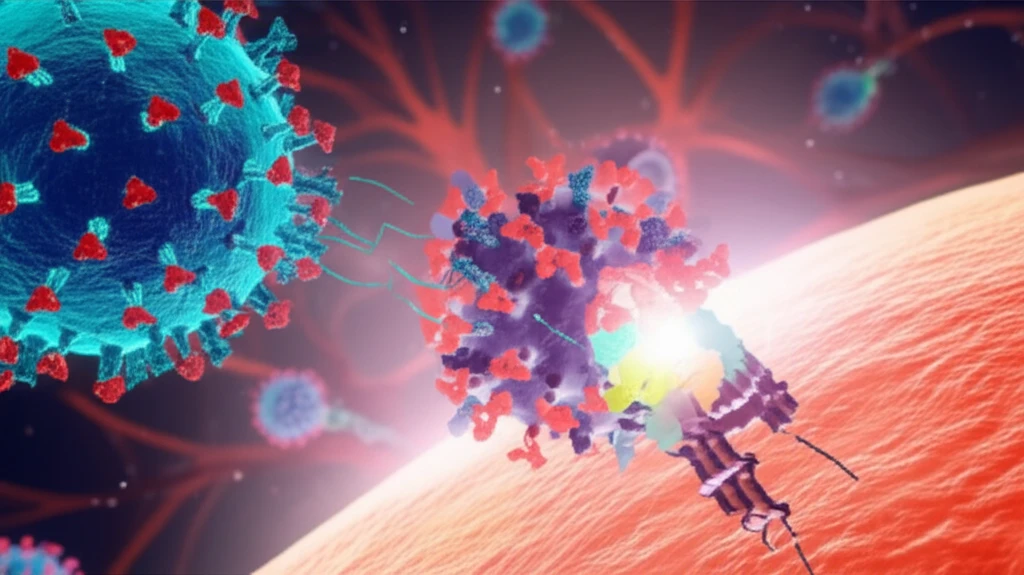
Decoding Measles: How a Tiny Protein Holds the Key to Stopping Infection
"Scientists uncover the secrets of the morbillivirus attachment protein, paving the way for better antiviral strategies."
Measles, a highly contagious viral disease, remains a significant global health concern despite the availability of an effective vaccine. Caused by the morbillivirus, measles can lead to severe complications, including pneumonia, encephalitis, and even death, particularly in young children and immunocompromised individuals. Understanding the intricate mechanisms by which the measles virus infects cells is crucial for developing targeted antiviral therapies and improving vaccination strategies.
The process of measles infection begins when the virus attaches to a host cell. This crucial step is mediated by the morbillivirus attachment protein, known as the H protein. The H protein is a complex structure that protrudes from the surface of the virus and binds to specific receptors on the surface of host cells. This interaction initiates a series of events that ultimately lead to the fusion of the viral membrane with the host cell membrane, allowing the virus to enter and replicate.
Recent research has focused on the structure and function of the H protein, aiming to identify key regions that are essential for its role in infection. One particular area of interest is the 'head-to-stalk linker' within the H protein. This linker region connects the receptor-binding head of the protein to its stalk, which anchors it to the viral membrane. Scientists believe that this linker plays a critical role in regulating the conformational changes necessary for membrane fusion, a vital step in the infection process.
Unlocking the Secrets of the Morbillivirus Attachment Protein

A groundbreaking study published in the Journal of Virology has provided new insights into the regulatory role of the morbillivirus attachment protein's head-to-stalk linker in membrane fusion triggering. Researchers have identified specific amino acids within this linker region that are critical for the protein's function, paving the way for the development of novel antiviral strategies targeting this essential step in the viral life cycle. The study focused on the role of isoleucine 146, revealing how its manipulation profoundly impacts viral entry.
- Mutagenesis Analysis: Researchers created a series of mutant H proteins, each with a different amino acid substitution in the head-to-stalk linker region.
- Cell-Based Assays: These mutant proteins were then tested in cell-based assays to assess their ability to bind to host cell receptors, promote membrane fusion, and facilitate viral entry.
- Structural Analysis: The team also used computational modeling to predict how these amino acid substitutions might affect the structure and dynamics of the H protein.
Future Directions and Potential Therapies
The results of this study have significant implications for the development of new antiviral therapies targeting the measles virus. By identifying key regions within the H protein that are essential for its function, researchers can now design drugs that specifically disrupt these regions, preventing the virus from infecting cells. One promising approach is to develop small molecules that bind to the head-to-stalk linker, interfering with its ability to regulate membrane fusion. Another strategy is to design antibodies that target the H protein, preventing it from binding to host cell receptors.
