Mouse model of liver cancer connected to glowing cellular networks

Decoding Liver Cancer: How Mouse Models are Revolutionizing Human Treatment

"Discover how C3HeB/FeJ mice are helping researchers unlock new insights into hepatocellular carcinoma and pave the way for better therapies."


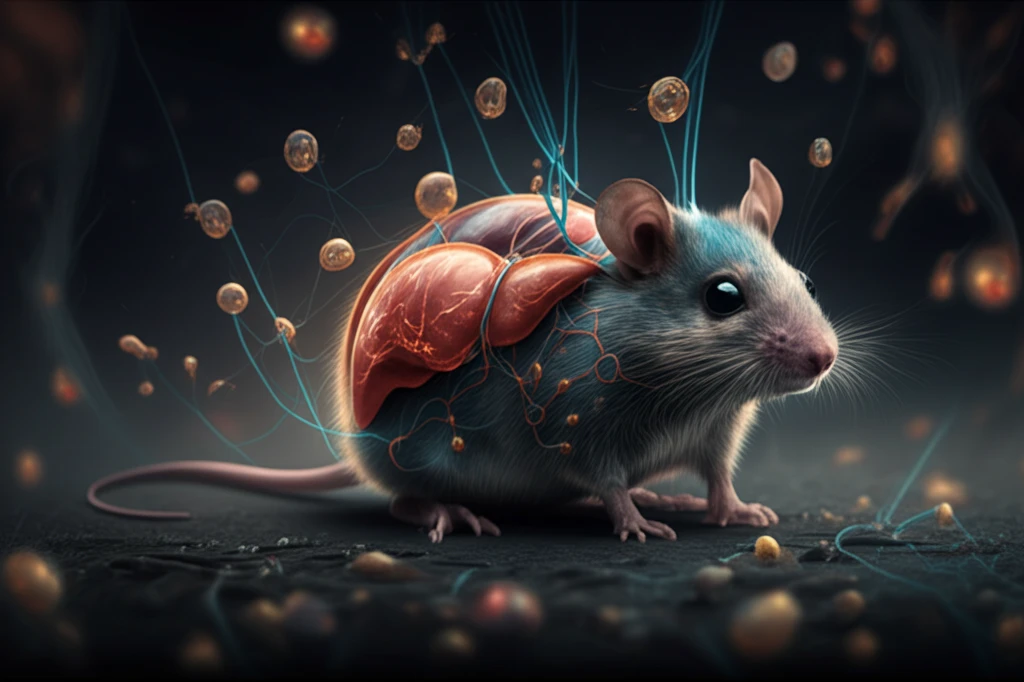
Hepatocellular carcinoma (HCC), a prevalent form of liver cancer, poses a significant global health challenge. Its insidious nature and resistance to treatment underscore the urgent need for robust preclinical models that accurately reflect the complexities of human disease. Traditional models often fall short, failing to capture the heterogeneity and nuanced progression of HCC in patients.

Enter the C3HeB/FeJ mouse, an inbred strain exhibiting spontaneous HCC development. Researchers have found that these mice naturally develop liver tumors that share striking similarities with human HCC, making them an invaluable asset for unraveling the disease's underlying mechanisms and testing novel therapeutic strategies. The spontaneous nature of tumor development in these mice mimics the unpredictable onset of human liver cancer.

This article explores how the C3HeB/FeJ mouse model is revolutionizing our understanding of HCC. From genetic parallels to advanced imaging techniques and gene expression analysis, we delve into the multifaceted ways these mice are helping scientists bridge the gap between preclinical research and clinical success. Discover how these findings translate into better diagnostic tools and more effective, personalized treatments for those battling liver cancer.

The C3HeB/FeJ Mouse Model: A Mirror for Human HCC

Mouse model of liver cancer connected to glowing cellular networks

The C3HeB/FeJ mouse model stands out due to its ability to spontaneously develop HCC, mirroring the natural onset of human liver cancer. These mice exhibit tumors with histological features remarkably similar to those found in human patients, including hepatosteatosis, dysplasia progression, vascular invasion, and diverse cell variants. Such similarities are crucial for accurately studying disease progression and therapeutic responses.

Researchers employ advanced diagnostic tools, such as ultrasound imaging, to monitor tumor development in C3HeB/FeJ mice. Ultrasound effectively detects and tracks tumors with high sensitivity, allowing for longitudinal studies of tumor growth and response to treatment. This non-invasive approach provides invaluable data on tumor behavior over time, mimicking clinical monitoring in human patients. Key findings include:

  • Tumors in C3HeB/FeJ mice exhibit hyperechogenicity with distinct borders, similar to human HCC.
  • Ultrasound imaging accurately correlates with physical tumor volume, providing a reliable non-invasive measurement.
  • Longitudinal monitoring allows researchers to observe diverse patterns of tumor response to therapy, including growth, regression, and delayed response.
Delving into the molecular landscape, gene expression analysis reveals significant overlap between C3HeB/FeJ mouse tumors and human HCC. Studies have identified thousands of differentially expressed genes shared between mouse and human tumors, particularly those involved in cancer pathways, cell cycle regulation, and p53 signaling. This molecular convergence reinforces the translational relevance of the C3HeB/FeJ model for studying human HCC.

Future Directions: Bridging the Gap to Human Therapies

The C3HeB/FeJ mouse model offers a powerful platform for preclinical research, accelerating the development of more effective therapies for human HCC. By closely mimicking the genetic, histological, and diagnostic features of human liver cancer, these mice enable researchers to identify novel drug targets, test therapeutic interventions, and refine treatment strategies. Future studies focused on personalized medicine approaches, leveraging the heterogeneity of C3HeB/FeJ tumors, promise to bring us closer to conquering this deadly disease.

About this Article -

This article was crafted using a human-AI hybrid and collaborative approach. AI assisted our team with initial drafting, research insights, identifying key questions, and image generation. Our human editors guided topic selection, defined the angle, structured the content, ensured factual accuracy and relevance, refined the tone, and conducted thorough editing to deliver helpful, high-quality information.See our About page for more information.

Everything You Need To Know

1

Why is the C3HeB/FeJ mouse model considered such a valuable tool in hepatocellular carcinoma (HCC) research?

The C3HeB/FeJ mouse model is significant because it spontaneously develops hepatocellular carcinoma (HCC) that closely mirrors the human form of the disease. These mice exhibit tumors with similar histological features to human HCC, including hepatosteatosis, dysplasia progression, vascular invasion, and diverse cell variants. This makes them a valuable tool for studying the progression of liver cancer and testing potential treatments.

2

How is ultrasound imaging used in the study of liver cancer within the C3HeB/FeJ mouse model, and what key findings have been revealed through its use?

Researchers utilize ultrasound imaging to monitor tumor development in C3HeB/FeJ mice. The tumors in these mice exhibit hyperechogenicity with distinct borders, much like human HCC. This non-invasive method allows for longitudinal studies, enabling researchers to observe how tumors respond to various therapies over time. This mimics clinical monitoring used for human patients.

3

What do gene expression analyses reveal about the similarities between tumors in C3HeB/FeJ mice and human hepatocellular carcinoma (HCC)?

Gene expression analysis has revealed significant overlap between C3HeB/FeJ mouse tumors and human HCC. Studies have identified thousands of differentially expressed genes shared between both, particularly in cancer pathways, cell cycle regulation, and p53 signaling. This genetic similarity strengthens the translational relevance of using C3HeB/FeJ mice to study human HCC and develop effective treatments.

4

In what ways does the C3HeB/FeJ mouse model facilitate the development of more effective therapies for human hepatocellular carcinoma (HCC)?

The use of the C3HeB/FeJ mouse model allows researchers to identify new drug targets and test different therapeutic interventions. By closely mimicking the genetic and histological features of human liver cancer, these mice help in refining treatment strategies. This model supports the exploration of personalized medicine approaches, leveraging the heterogeneity of tumors found in C3HeB/FeJ mice to tailor treatments more effectively.

5

What are the future directions for research using the C3HeB/FeJ mouse model, and how might they improve outcomes for patients with hepatocellular carcinoma (HCC)?

Future research using the C3HeB/FeJ mouse model will focus on creating personalized medicine approaches to treat hepatocellular carcinoma (HCC). Given the diversity of tumors that develop in these mice, researchers can refine therapies to target specific genetic and molecular characteristics of individual tumors. This could lead to more effective and tailored treatments for patients battling liver cancer. It's important to also consider other HCC models available, as their strengths can complement the C3HeB/FeJ model.

Newsletter Subscribe

Subscribe to get the latest articles and insights directly in your inbox.