Illustration of a brain with neural pathways and DNA strand, representing glioma treatment advancements.

Decoding Glioma: How Advanced Imaging and Molecular Analysis are Changing Brain Tumor Treatment

"Explore the latest research on gliomas, from predicting treatment response with radiomics to understanding contrast enhancement through molecular physiology, and how these advancements pave the way for personalized therapies."


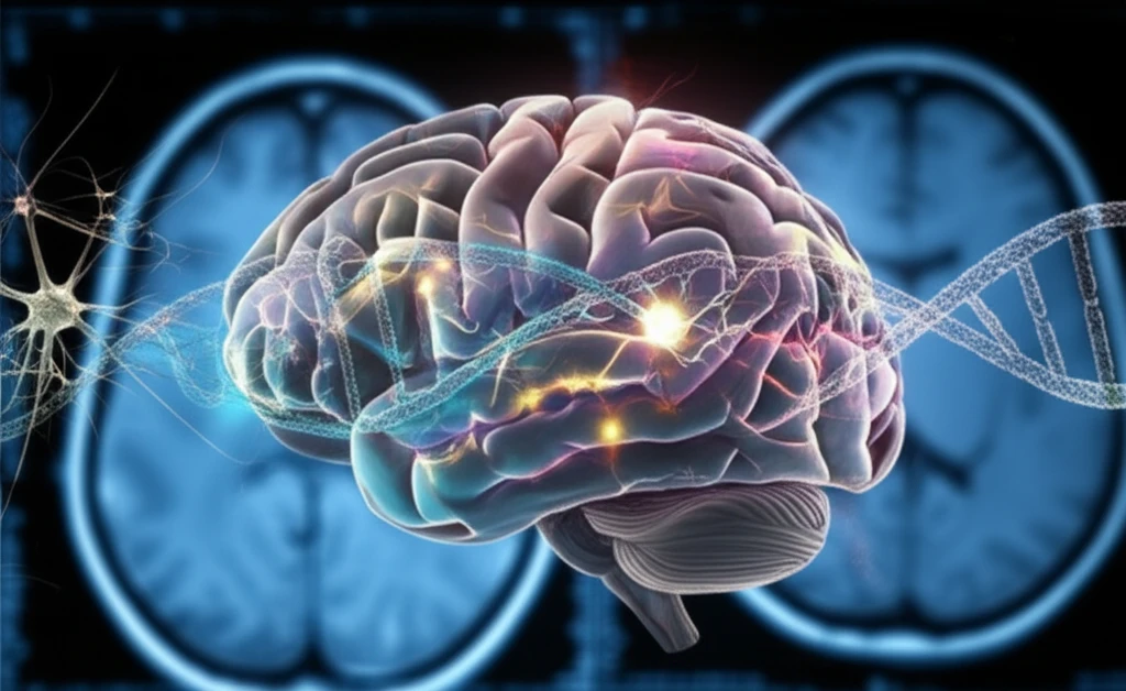
Glioblastomas and other high-grade gliomas represent a significant challenge in oncology due to their aggressive nature and resistance to treatment. Recent research is focusing on leveraging advanced imaging techniques and molecular analysis to better understand these tumors and improve patient outcomes. This article explores several key studies presented at a recent neuro-oncology conference, shedding light on the latest advancements in glioma research.

One promising area of research involves the use of diffusion MRI to identify early signs of tumor progression in lower-grade gliomas. By tracking changes in diffusion parameters, clinicians may be able to detect progression before it is visible through traditional imaging methods, allowing for earlier intervention.

Another focus is on understanding the molecular physiology of contrast enhancement in glioblastomas. Contrast enhancement on MRI is a key feature used to assess these tumors, but the underlying biology is complex. By analyzing the relationship between contrast enhancement and gene expression, researchers are gaining insights into the processes that drive tumor growth and response to therapy.

Predicting Treatment Response with Radiomics and Genomic Data

Illustration of a brain with neural pathways and DNA strand, representing glioma treatment advancements.

Radiomics, the process of extracting quantitative features from medical images, is emerging as a powerful tool for predicting treatment response in glioblastomas. A recent study explored the use of radiomic features from pre-treatment MRI scans to predict response to chemo-radiation therapy. The study found that specific radiomic features, particularly those related to texture and heterogeneity, were predictive of response. These features were also correlated with key signaling pathways involved in treatment resistance, such as the AKT pathway.

Specifically, Laws energy features, which characterize the appearance of ripples and spots in MRI images, were found to be strong predictors of response. These features correlated significantly with AKT and apoptosis signaling pathways. Activation of the AKT pathway is known to promote angiogenesis and resistance to chemo-radiation, while apoptosis is a process of programmed cell death that can be triggered by treatment. Tumors with higher expression of apoptotic proteins tend to respond better to therapy.

  • Study Details: Researchers analyzed MRI scans and RNA sequencing data from glioblastoma patients to identify radiomic features associated with treatment response.
  • Key Findings: Radiomic features from pre-treatment MRI scans were predictive of response to chemo-radiation therapy.
  • Biological Correlation: Predictive radiomic features were correlated with AKT and apoptosis signaling pathways.
  • Clinical Implication: Radiomics may allow for non-invasive prediction of treatment response, helping to guide treatment decisions.
Another study delved into radiogenomic analysis to correlate textural features from MRI with genomic immune scores. The goal was to find imaging markers that reflect the level of immune response within the tumor, as a strong immune response is generally associated with better outcomes in glioblastoma patients treated with chemo-radiation therapy. The researchers discovered that specific textural features on MRI were indeed correlated with immune scores derived from gene expression data. These features captured wave-like enhancement patterns and intensity heterogeneity within the tumor and surrounding edema.

The Future of Glioma Treatment

The research discussed here highlights the potential of advanced imaging and molecular analysis to transform the treatment of gliomas. By integrating radiomics, genomics, and other data, clinicians can gain a more comprehensive understanding of each patient's tumor and tailor treatment accordingly. This personalized approach promises to improve outcomes and quality of life for individuals affected by these challenging cancers.

About this Article -

This article was crafted using a human-AI hybrid and collaborative approach. AI assisted our team with initial drafting, research insights, identifying key questions, and image generation. Our human editors guided topic selection, defined the angle, structured the content, ensured factual accuracy and relevance, refined the tone, and conducted thorough editing to deliver helpful, high-quality information.See our About page for more information.

Everything You Need To Know

1

What are the main challenges in treating gliomas, and how is research addressing these challenges?

Glioblastomas and other high-grade gliomas pose a significant challenge in oncology due to their aggressive nature and resistance to treatment. Researchers are using advanced imaging techniques and molecular analysis to better understand these tumors and improve patient outcomes. This work is important because it seeks to move beyond generic treatment approaches toward tailored interventions based on the unique characteristics of each patient's tumor. The aggressive nature and resistance to treatment mean that novel approaches are needed to improve outcomes and quality of life for individuals affected by these challenging cancers.

2

How can Diffusion MRI be used in the treatment of gliomas?

Diffusion MRI helps clinicians detect early signs of tumor progression in lower-grade gliomas. By tracking changes in diffusion parameters, clinicians can potentially identify progression before it's visible through traditional methods, which allows for earlier intervention. Traditional imaging methods might not always catch these subtle changes early on. Early detection enables timely intervention, potentially improving the effectiveness of treatments and patient outcomes. This shift from reactive to proactive care represents a significant advancement in the management of gliomas.

3

What is radiomics, and how can it help in treating gliomas?

Radiomics involves extracting quantitative features from medical images, emerging as a tool for predicting treatment response in glioblastomas. One study used radiomic features from pre-treatment MRI scans to predict response to chemo-radiation therapy. Specific radiomic features, like Laws energy features that characterize the appearance of ripples and spots in MRI images were found to be strong predictors of response, and were correlated with the AKT and apoptosis signaling pathways. This is important because it allows for non-invasive prediction of treatment response, which can help guide treatment decisions. This personalized approach promises to improve outcomes.

4

Why is understanding contrast enhancement on MRI important in the context of brain tumor treatment?

Contrast enhancement on MRI is a key feature used to assess glioblastomas, but the underlying biology is complex. Researchers analyze the relationship between contrast enhancement and gene expression. Understanding the molecular physiology of contrast enhancement helps researchers understand the tumor’s behavior and response to therapy. By understanding the biological processes behind contrast enhancement, clinicians can gain insights into tumor growth and tailor treatment accordingly. This approach moves towards more effective, targeted therapies.

5

How does radiogenomic analysis contribute to understanding and treating gliomas?

Radiogenomic analysis correlates textural features from MRI with genomic immune scores to find imaging markers reflecting the immune response within the tumor. The research discovered that specific textural features on MRI correlated with immune scores derived from gene expression data. These features captured wave-like enhancement patterns and intensity heterogeneity within the tumor and surrounding edema. A strong immune response is generally associated with better outcomes in glioblastoma patients treated with chemo-radiation therapy. This is crucial because a robust immune response often correlates with improved treatment outcomes, thus allowing for personalized therapies.

Newsletter Subscribe

Subscribe to get the latest articles and insights directly in your inbox.