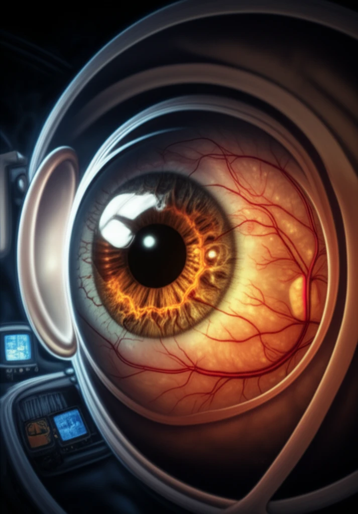
Advanced retina scan with fractal patterns, symbolizing detailed eye health analysis.

Decoding Diabetic Retinopathy: How Advanced Eye Scans Can Save Your Sight

"New fractal and lacunarity analyses offer a more detailed way to assess microvascular damage, leading to earlier and more accurate diagnosis."


Diabetic retinopathy (DR) remains a leading cause of blindness worldwide, impacting millions of working-age adults. This condition arises from chronic high blood sugar levels, which damage the tiny blood vessels in the retina. Early detection and management are crucial to preventing severe vision loss, making regular eye exams a non-negotiable part of diabetes care.

Traditional methods of screening for diabetic retinopathy, such as fundus photography and fluorescein angiography, have limitations. While effective for detecting advanced stages of the disease, they often miss subtle changes in the microvasculature that occur in the early stages. This is where newer technologies like Optical Coherence Tomography Angiography (OCTA) come into play, offering a more detailed and non-invasive way to visualize retinal blood vessels.

OCTA provides high-resolution images of the retinal microvasculature, allowing doctors to see individual capillary layers. This technology opens new possibilities for quantifying and analyzing the intricate patterns of blood vessels in the eye. Recent research has focused on using advanced mathematical techniques, such as fractal and lacunarity analyses, to extract even more information from OCTA images. These methods promise to improve our ability to diagnose DR earlier and more accurately, paving the way for more effective treatments.

What Are Fractal and Lacunarity Analyses, and Why Do They Matter for Diabetic Retinopathy?

Advanced retina scan with fractal patterns, symbolizing detailed eye health analysis.

Fractal and lacunarity analyses are sophisticated mathematical techniques used to characterize complex patterns. In the context of diabetic retinopathy, these methods help to quantify subtle changes in the structure of retinal blood vessels that are often invisible to the naked eye. By applying these analyses to OCTA images, researchers can gain deeper insights into the microvascular damage caused by diabetes.

Fractal analysis assesses the self-similarity and scaling independence of vascular networks. This means it looks at how similar the patterns are at different magnifications. In healthy retinas, blood vessels exhibit a certain degree of fractal complexity. However, in diabetic retinopathy, this complexity is often disrupted due to vessel damage and loss.

  • Self-Similarity: Examines how similar the vascular patterns are at different scales.
  • Scaling Independence: Determines if the patterns maintain their characteristics regardless of magnification.
  • Complexity Disruption: Identifies how diabetes alters the natural fractal complexity of retinal vessels.
Lacunarity analysis, on the other hand, measures the size and distribution of gaps or voids within the vascular network. It provides information about the texture and homogeneity of the retinal vessels. In diabetic retinopathy, increased lacunarity can indicate areas of capillary dropout and non-perfusion, reflecting the severity of microvascular damage. Together, fractal and lacunarity analyses offer a comprehensive assessment of retinal microvascular health, providing valuable information for early diagnosis and disease management.

Empowering Patients Through Advanced Diagnostics

The integration of fractal and lacunarity analyses with OCTA imaging represents a significant advancement in the diagnosis and management of diabetic retinopathy. These sophisticated techniques provide a more detailed and quantitative assessment of retinal microvascular health, enabling earlier detection of subtle changes indicative of DR. By empowering clinicians with this enhanced diagnostic capability, we can move closer to preventing vision loss and improving the quality of life for individuals with diabetes. Regular eye exams incorporating these advanced methods are essential for proactive and effective diabetes care.

About this Article -

This article was crafted using a human-AI hybrid and collaborative approach. AI assisted our team with initial drafting, research insights, identifying key questions, and image generation. Our human editors guided topic selection, defined the angle, structured the content, ensured factual accuracy and relevance, refined the tone, and conducted thorough editing to deliver helpful, high-quality information.See our About page for more information.

This article is based on research published under:

DOI-LINK: 10.1111/micc.12519, Alternate LINK

Title: Multifractal And Lacunarity Analyses Of Microvascular Morphology In Eyes With Diabetic Retinopathy: A Projection Artifact Resolved Optical Coherence Tomography Angiography Study

Subject: Physiology (medical)

Journal: Microcirculation

Publisher: Wiley

Authors: Tiepei Zhu, Jinlian Ma, Jinyu Li, Xizhe Dai, Panpan Ye, Zhaoan Su, Dexing Kong, Juan Ye

Published: 2019-03-20

Everything You Need To Know

1

What is diabetic retinopathy, and why should I be concerned about it if I have diabetes?

Diabetic retinopathy is a condition that results from damage to the tiny blood vessels in the retina due to chronic high blood sugar levels, frequently impacting working-age adults and is a leading cause of blindness worldwide. Early detection is crucial, which makes regular eye exams essential for those with diabetes. While fundus photography and fluorescein angiography have been used, they have limitations in detecting early changes. Newer technologies like Optical Coherence Tomography Angiography (OCTA) offer detailed views of retinal blood vessels, allowing for a more in-depth analysis.

2

What is Optical Coherence Tomography Angiography (OCTA), and why is it important in diagnosing diabetic retinopathy?

Optical Coherence Tomography Angiography, or OCTA, is significant because it provides high-resolution images of the retinal microvasculature, allowing doctors to visualize individual capillary layers. This technology enables the quantification and analysis of intricate blood vessel patterns in the eye. Combined with advanced mathematical techniques such as fractal and lacunarity analyses, OCTA enhances the ability to diagnose diabetic retinopathy earlier and more accurately, leading to more effective treatments and potential prevention of vision loss.

3

Can you explain what fractal analysis is and how it is used to detect diabetic retinopathy?

Fractal analysis assesses the self-similarity and scaling independence of vascular networks within the retina. It examines how similar vascular patterns are at different magnifications and determines if these patterns maintain their characteristics regardless of magnification. In healthy retinas, blood vessels exhibit fractal complexity. However, in diabetic retinopathy, this complexity is often disrupted due to vessel damage and loss. Fractal analysis helps identify how diabetes alters the natural fractal complexity of retinal vessels, offering insights into the progression of the disease.

4

What does lacunarity analysis measure, and how does it help in assessing diabetic retinopathy?

Lacunarity analysis measures the size and distribution of gaps or voids within the retinal vascular network. It provides information about the texture and homogeneity of retinal vessels. In the context of diabetic retinopathy, increased lacunarity can indicate areas of capillary dropout and non-perfusion, reflecting the severity of microvascular damage. Unlike fractal analysis, which focuses on the complexity of vessel patterns, lacunarity analysis specifically targets the irregularities and spaces within the vascular structure, providing a complementary perspective on retinal health.

5

How do fractal and lacunarity analyses together improve the diagnosis and management of diabetic retinopathy?

The integration of fractal and lacunarity analyses with OCTA imaging enhances diagnostic capabilities by providing a more detailed and quantitative assessment of retinal microvascular health. This combination allows for earlier detection of subtle changes indicative of diabetic retinopathy, empowering clinicians to prevent vision loss and improve the quality of life for individuals with diabetes. Regular eye exams that incorporate these advanced methods are essential for proactive and effective diabetes care, ensuring that early signs of damage are identified and addressed promptly.

Newsletter Subscribe

Subscribe to get the latest articles and insights directly in your inbox.