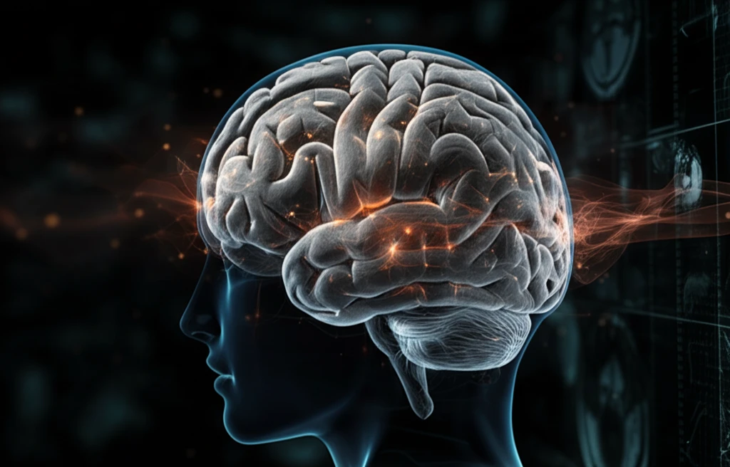
AI texture analysis of a brain MRI scan.

Decoding Brain Scans: Can AI Texture Analysis Improve Glioma Diagnosis?

"Explore how AI-driven texture analysis enhances MRI interpretation for glioma classification, potentially revolutionizing brain tumor diagnostics for better patient outcomes."


Gliomas, tumors originating in the brain's glial cells, present a significant diagnostic challenge due to their varied nature. Accurate classification of these tumors is crucial as it directly impacts treatment strategies and patient prognosis. Traditionally, this classification relies on histopathological examination and genetic analysis, coupled with the interpretation of Magnetic Resonance Imaging (MRI) scans.

However, MRI interpretation can be subjective and may miss subtle yet critical details. This is where texture analysis steps in—a sophisticated method that uses algorithms to quantify the textural patterns within an image that might be imperceptible to the human eye. Recent advancements have seen the integration of artificial intelligence (AI) to enhance this analysis, promising a more objective and accurate diagnostic process.

This article delves into how AI-enhanced texture analysis is transforming the diagnostic landscape for gliomas. By examining a recent study that explores the application of AI in classifying low-grade gliomas (LGGs) using MRI data, we shed light on the potential benefits of this innovative approach.

How Does Texture Analysis Work in Glioma Diagnosis?

AI texture analysis of a brain MRI scan.

Texture analysis works by examining the spatial relationships of pixel intensities in an MRI image. In the context of glioma diagnosis, it helps to identify and quantify characteristics such as homogeneity, contrast, and structural arrangements within the tumor. These features can then be correlated with different grades and types of gliomas, providing valuable diagnostic insights.

The recent study focused on using texture analysis to differentiate between astrocytomas and oligodendrogliomas—two common types of LGGs. The researchers applied texture analysis to conventional MRI sequences and identified key texture features that could accurately distinguish between these tumor types.

  • Data Collection: MRI scans from 41 patients with LGGs were used, including both astrocytomas and oligodendrogliomas.
  • Image Analysis: Texture analysis was performed on various MRI sequences, including T2-weighted FLAIR, ADC, T1-weighted, and T1-weighted with gadolinium.
  • Feature Extraction: The analysis extracted 279 texture features from each MRI image, encompassing statistical measures like histogram features, gradient features, and run-length matrices.
  • AI Integration: Machine learning algorithms were employed to identify the most discriminant features, enhancing the accuracy of tumor classification.
Key to the success of this method is the use of machine learning algorithms to filter and prioritize the most relevant texture features. These algorithms help in reducing the complexity of the data and focusing on the markers that are most indicative of specific tumor types. This AI-driven approach not only improves accuracy but also enhances the efficiency of the diagnostic process.

The Future of Glioma Diagnostics with AI

The integration of AI into texture analysis represents a significant leap forward in the diagnosis and classification of gliomas. By providing a more objective, accurate, and efficient method for analyzing MRI data, this technology has the potential to improve patient outcomes through better-informed treatment decisions. As AI continues to evolve, its role in medical imaging and diagnostics will likely expand, paving the way for more personalized and effective healthcare strategies.

About this Article -

This article was crafted using a human-AI hybrid and collaborative approach. AI assisted our team with initial drafting, research insights, identifying key questions, and image generation. Our human editors guided topic selection, defined the angle, structured the content, ensured factual accuracy and relevance, refined the tone, and conducted thorough editing to deliver helpful, high-quality information.See our About page for more information.

This article is based on research published under:

DOI-LINK: 10.1016/j.mri.2018.11.008, Alternate LINK

Title: Mri Based Texture Analysis To Classify Low Grade Gliomas Into Astrocytoma And 1P/19Q Codeleted Oligodendroglioma

Subject: Radiology, Nuclear Medicine and imaging

Journal: Magnetic Resonance Imaging

Publisher: Elsevier BV

Authors: Shun Zhang, Gloria Chia-Yi Chiang, Rajiv S. Magge, Howard Alan Fine, Rohan Ramakrishna, Eileen Wang Chang, Tejas Pulisetty, Yi Wang, Wenzhen Zhu, Ilhami Kovanlikaya

Published: 2019-04-01

Everything You Need To Know

1

How does texture analysis work to improve glioma diagnosis, and what specific features does it evaluate in MRI images?

Texture analysis, especially when enhanced by AI, examines MRI images by quantifying textural patterns. It identifies and measures characteristics like homogeneity, contrast, and structural arrangements within a potential glioma. These features are then correlated with different grades and types of gliomas, offering diagnostic insights that might be missed by visual inspection alone. This approach is particularly valuable for distinguishing between different types of low-grade gliomas (LGGs).

2

What are the key steps involved in using AI-enhanced texture analysis for glioma diagnosis, starting from data collection to final classification?

The diagnostic process leverages MRI scans to collect data. Texture analysis is performed to extract 279 texture features, including histogram features, gradient features, and run-length matrices. Machine learning algorithms then identify the most discriminant features to enhance tumor classification accuracy. This process helps differentiate between tumor types, such as astrocytomas and oligodendrogliomas.

3

In what specific ways does AI integration enhance the accuracy and efficiency of texture analysis in distinguishing between different types of gliomas?

AI integration refines texture analysis by prioritizing the most relevant texture features using machine learning algorithms. These algorithms reduce data complexity, focusing on markers indicative of specific tumor types. This AI-driven approach improves diagnostic accuracy and enhances the efficiency of the overall process, reducing the potential for human error.

4

What are the potential benefits of using AI-enhanced texture analysis in glioma diagnostics for patient outcomes and treatment strategies?

The application of AI-enhanced texture analysis can lead to improved patient outcomes through more accurate and timely diagnoses of gliomas. By providing a more objective and efficient method for analyzing MRI data, clinicians can make better-informed treatment decisions. This approach has the potential to personalize healthcare strategies, ensuring patients receive treatments tailored to the specific characteristics of their tumor.

5

What are the limitations of current AI-enhanced texture analysis methods for glioma diagnosis, and what future research directions could address these limitations?

While the study successfully used AI-enhanced texture analysis to differentiate between astrocytomas and oligodendrogliomas, it's important to remember that gliomas are incredibly varied. Future research needs to investigate the application of this method across a broader spectrum of glioma types and grades. Moreover, integrating genetic analysis with texture analysis could further refine diagnostic accuracy. Additionally, exploring the utility of other advanced imaging techniques beyond conventional MRI could improve the AI models used in the texture analysis.

Newsletter Subscribe

Subscribe to get the latest articles and insights directly in your inbox.