Leukemia cells transforming into healthy blood cells

Decoding AML M3: A Comprehensive Guide to Acute Promyelocytic Leukemia

"Navigate the complexities of Acute Promyelocytic Leukemia (APL) with our in-depth review, covering diagnosis, treatment, and the latest genetic insights."


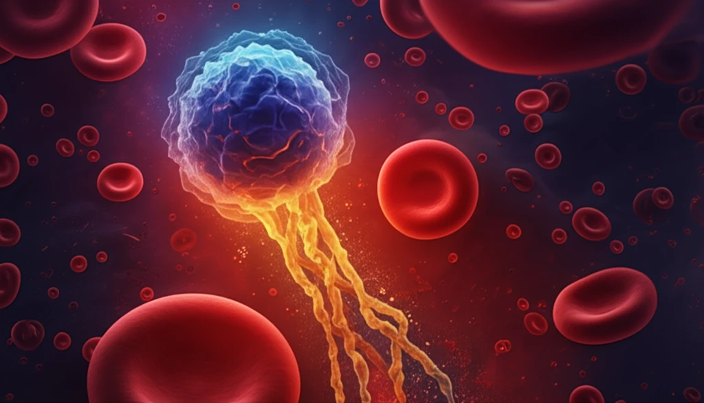
Acute Promyelocytic Leukemia (APL), a distinct subtype of acute myeloid leukemia (AML), is characterized by unique clinical and molecular features. Understanding APL is crucial for effective diagnosis and treatment, which have dramatically improved in recent years. This article provides a comprehensive review of APL, covering its subtypes, genetic underpinnings, diagnostic criteria, and current therapeutic strategies.

APL is specifically defined by the presence of abnormal promyelocytes in the bone marrow, often accompanied by a specific chromosomal translocation involving the retinoic acid receptor alpha (RARA) gene on chromosome 17. The most common translocation, t(15;17)(q24;q21), results in the fusion of the promyelocytic leukemia (PML) gene on chromosome 15 with the RARA gene. This PML/RARA fusion protein plays a key role in the pathogenesis of APL.

Historically, APL was associated with high mortality due to bleeding complications, but the introduction of all-trans retinoic acid (ATRA) and arsenic trioxide (ATO) has revolutionized treatment. These agents induce differentiation of leukemic cells and have significantly improved survival rates, making APL one of the most curable forms of acute leukemia.

Understanding APL Subtypes and Genetic Variations

Leukemia cells transforming into healthy blood cells

APL is primarily classified into two main subtypes based on morphology: the typical or hypergranular form (AML M3) and the microgranular variant (AML M3v). AML M3 is characterized by promyelocytes with abundant granules and Auer rods, which are crystalline aggregates of myeloperoxidase. In contrast, AML M3v exhibits promyelocytes with minimal granulation and bilobed nuclei. The microgranular variant often presents with a higher white blood cell count and a greater risk of disseminated intravascular coagulation (DIC).

While the PML/RARA translocation is the hallmark of APL, variant translocations involving RARA with other partner genes have been identified in a small percentage of cases. These variants, such as those involving ZBTB16 (PLZF), NUMA1, or NPM1, may have different clinical characteristics and responses to treatment. Identifying these variant translocations is important for tailoring therapy and predicting prognosis.

Key genetic features in APL include:
  • PML/RARA fusion from t(15;17)(q24;q21)
  • Variant translocations involving RARA with ZBTB16, NUMA1, or NPM1
  • Additional chromosomal abnormalities (ACA) in some cases
Diagnostic evaluation of APL involves a combination of morphological assessment, cytogenetic analysis, and molecular testing. Bone marrow examination is essential for identifying abnormal promyelocytes and assessing their morphology. Cytogenetic analysis, including karyotyping and fluorescence in situ hybridization (FISH), is used to detect the PML/RARA translocation or variant translocations. Molecular testing, such as reverse transcription-polymerase chain reaction (RT-PCR), is highly sensitive for detecting PML/RARA transcripts and monitoring response to treatment.

Advances in APL Treatment and Monitoring

The treatment of APL has evolved significantly with the introduction of ATRA and ATO, which have replaced traditional chemotherapy-based regimens in many cases. ATRA induces differentiation of promyelocytes, while ATO promotes apoptosis. Combination therapy with ATRA and ATO has shown remarkable efficacy, leading to high rates of complete remission and long-term survival, particularly in low- to intermediate-risk patients. Regular monitoring with RQ-PCR is crucial for detecting minimal residual disease and preventing relapse.

About this Article -

This article was crafted using a human-AI hybrid and collaborative approach. AI assisted our team with initial drafting, research insights, identifying key questions, and image generation. Our human editors guided topic selection, defined the angle, structured the content, ensured factual accuracy and relevance, refined the tone, and conducted thorough editing to deliver helpful, high-quality information.See our About page for more information.

Everything You Need To Know

1

What genetic translocation is most commonly associated with Acute Promyelocytic Leukemia (APL), and what genes are involved?

The most common genetic translocation associated with Acute Promyelocytic Leukemia (APL) is t(15;17)(q24;q21). This translocation results in the fusion of the promyelocytic leukemia (PML) gene on chromosome 15 with the retinoic acid receptor alpha (RARA) gene on chromosome 17, creating the PML/RARA fusion protein. The PML/RARA fusion protein is a key factor in the development and progression of APL by blocking myeloid differentiation. While this translocation is the most prevalent, variant translocations involving RARA with other genes such as ZBTB16, NUMA1, or NPM1 can also occur, though less frequently. These variants might influence clinical characteristics and responses to treatment.

2

How have treatments like ATRA and arsenic trioxide (ATO) changed the prognosis for individuals diagnosed with Acute Promyelocytic Leukemia (APL)?

The introduction of all-trans retinoic acid (ATRA) and arsenic trioxide (ATO) has revolutionized the treatment of Acute Promyelocytic Leukemia (APL). Historically, APL was associated with high mortality rates due to bleeding complications. ATRA induces differentiation of leukemic promyelocytes, while ATO promotes apoptosis. These agents have significantly improved survival rates, making APL one of the most curable forms of acute leukemia. This combination therapy has shown remarkable efficacy, leading to high rates of complete remission and long-term survival, particularly in low- to intermediate-risk patients. The shift towards using ATRA and ATO has reduced the reliance on traditional chemotherapy-based regimens, which were more toxic and less effective.

3

What are the key differences between the typical (AML M3) and microgranular variant (AML M3v) subtypes of Acute Promyelocytic Leukemia (APL)?

The two main subtypes of Acute Promyelocytic Leukemia (APL) are the typical or hypergranular form (AML M3) and the microgranular variant (AML M3v). AML M3 is characterized by promyelocytes containing abundant granules and Auer rods. Auer rods are crystalline aggregates of myeloperoxidase. In contrast, AML M3v exhibits promyelocytes with minimal granulation and bilobed nuclei. The microgranular variant (AML M3v) often presents with a higher white blood cell count and a greater risk of disseminated intravascular coagulation (DIC). These morphological distinctions are crucial for accurate diagnosis and risk stratification, guiding appropriate treatment strategies for each subtype.

4

Beyond the standard PML/RARA fusion, what other genetic variations can occur in Acute Promyelocytic Leukemia (APL), and why is identifying them important?

While the PML/RARA fusion resulting from the t(15;17) translocation is the most common genetic feature in Acute Promyelocytic Leukemia (APL), variant translocations involving the retinoic acid receptor alpha (RARA) gene with other partner genes can occur. These include fusions with genes such as ZBTB16 (PLZF), NUMA1, and NPM1. Identifying these variant translocations is crucial because they can be associated with different clinical characteristics and responses to treatment. For example, some variants may not respond as well to standard all-trans retinoic acid (ATRA) therapy, necessitating alternative or more intensive treatment approaches. Therefore, detecting these variants is essential for tailoring therapy and predicting prognosis in APL patients.

5

How is minimal residual disease monitored in Acute Promyelocytic Leukemia (APL) after treatment, and why is this monitoring important for long-term outcomes?

In Acute Promyelocytic Leukemia (APL), minimal residual disease (MRD) is monitored primarily using reverse transcription-quantitative polymerase chain reaction (RQ-PCR). This technique detects PML/RARA transcripts, indicating the presence of residual leukemic cells. Regular monitoring with RQ-PCR is crucial for detecting minimal residual disease and preventing relapse. If MRD is detected, it may indicate the need for further intervention, such as consolidation therapy or stem cell transplantation, to eradicate the remaining leukemic cells and improve long-term survival. Consistent and sensitive MRD monitoring allows for timely intervention and helps to maintain high rates of complete remission and overall survival in APL patients.

Newsletter Subscribe

Subscribe to get the latest articles and insights directly in your inbox.