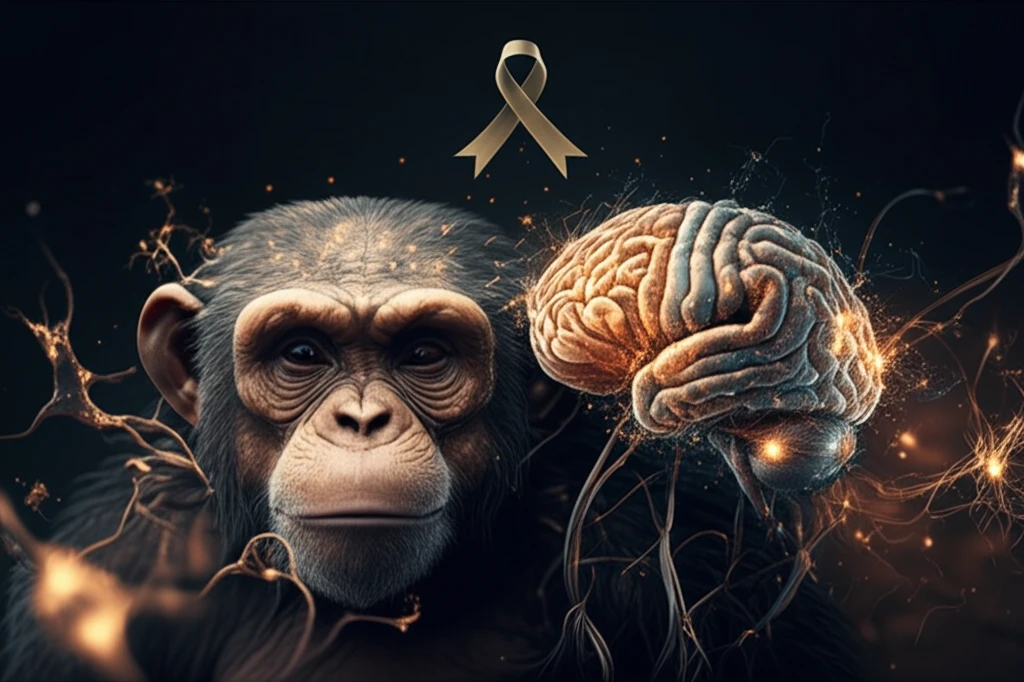
Chimpanzee and human brains intertwined, highlighting astrocytic differences in Alzheimer's research

Cracking the Aging Code: What Chimpanzee Brains Reveal About Alzheimer's

"New research explores how astrocytes in chimpanzee brains respond to aging and Alzheimer's-like pathology, offering surprising clues for human treatments"


Alzheimer's disease (AD) remains one of the most formidable challenges in modern medicine. As the global population ages, the prevalence of AD continues to rise, placing immense strain on healthcare systems and affecting millions of lives. While extensive research has been dedicated to understanding the mechanisms of AD, effective treatments and preventative strategies remain elusive.

Astrocytes, the most abundant glial cells in the brain, play a crucial role in maintaining the central nervous system's (CNS) health. These cells are involved in everything from ion and water balance to neurotransmitter regulation and synaptic modulation. However, in the context of AD, astrocytes can become reactive, leading to a state known as astrogliosis, marked by increased expression of glial fibrillary acidic protein (GFAP) and cellular hypertrophy. Understanding how astrocytes behave in aging and AD is critical for developing targeted therapies.

To shed light on these complex processes, a recent study investigated astrocytic changes in aging chimpanzees, our closest living relatives. Chimpanzees, like humans, can develop AD-type pathology, including amyloid plaques and neurofibrillary tangles, making them a valuable model for studying the disease. By comparing astrocytic changes in chimpanzees and humans, researchers hope to uncover unique insights that could lead to new therapeutic strategies for AD.

Astrocytes in Aging Chimpanzees: A Unique Perspective

Chimpanzee and human brains intertwined, highlighting astrocytic differences in Alzheimer's research

The study, conducted by Munger et al. (2019), examined the brains of 27 chimpanzees, ranging in age from 12 to 62 years. The researchers focused on specific brain regions, including the hippocampus (HC), mid-temporal gyrus (MTG), and prefrontal cortex (PFC), which are known to be affected by AD in humans. Using stereologic methods, they quantified GFAP-immunoreactive astrocyte density and soma volume in different cortical layers and hippocampal fields.

One of the most striking findings was that, unlike humans, GFAP expression in chimpanzees did not increase with age. This suggests that chimpanzees may experience lower oxidative stress loads or possess protective mechanisms that make their astrocytes less reactive to age-related changes. However, the study did reveal that chimpanzee layer I astrocytes in the PFC are susceptible to AD-like changes, similar to what is observed in humans.

The researchers identified several key differences in astrocytic behavior between chimpanzees and humans:
  • GFAP expression does not increase with age in chimpanzees.
  • Chimpanzee layer I astrocytes in the PFC are susceptible to AD-like changes, similar to humans.
  • Astrogliosis in chimpanzees is primarily restricted to layer I of the PFC.
  • Astrogliosis in the CA1 and CA3 subfields was associated with neuritic cluster density and amyloid plaque volume.
Further analysis revealed that astrogliosis in the CA1 and CA3 subfields was associated with neuritic cluster density and amyloid plaque volume. This suggests that while chimpanzees do not exhibit a global increase in astrogliosis with age, their astrocytes do respond to the presence of AD-type pathology in specific brain regions. Moreover, the study found that astrogliosis in chimpanzees was primarily restricted to layer I of the PFC, indicating a unique pattern of cortical aging in these primates.

Implications for Human Alzheimer's Treatment

These findings have significant implications for our understanding of AD in humans. By identifying the unique characteristics of astrocytic responses in chimpanzees, researchers can gain valuable insights into the complex interplay between aging, AD-type pathology, and astrocyte activation. This knowledge could pave the way for the development of targeted therapies that modulate astrocytic behavior to mitigate the progression of AD. Further research is needed to fully elucidate the mechanisms underlying these differences and to determine how they can be harnessed to improve human brain health.

About this Article -

This article was crafted using a human-AI hybrid and collaborative approach. AI assisted our team with initial drafting, research insights, identifying key questions, and image generation. Our human editors guided topic selection, defined the angle, structured the content, ensured factual accuracy and relevance, refined the tone, and conducted thorough editing to deliver helpful, high-quality information.See our About page for more information.

This article is based on research published under:

DOI-LINK: 10.1002/cne.24610, Alternate LINK

Title: Astrocytic Changes With Aging And Alzheimer'S Disease‐Type Pathology In Chimpanzees

Subject: General Neuroscience

Journal: Journal of Comparative Neurology

Publisher: Wiley

Authors: Emily L. Munger, Melissa K. Edler, William D. Hopkins, John J. Ely, Joseph M. Erwin, Daniel P. Perl, Elliott J. Mufson, Patrick R. Hof, Chet C. Sherwood, Mary Ann Raghanti

Published: 2019-01-07

Everything You Need To Know

1

What did the study reveal about GFAP expression in aging chimpanzees compared to humans?

The research indicates that unlike in humans, GFAP expression in chimpanzees does not increase with age. This suggests potential protective mechanisms in chimpanzees that make their astrocytes less reactive to age-related changes, possibly due to lower oxidative stress. However, a key finding is that chimpanzee layer I astrocytes in the PFC are susceptible to AD-like changes, similar to what is observed in humans.

2

What role do astrocytes play in the brain, and how does their behavior change in the context of Alzheimer's disease?

Astrocytes play crucial roles in the central nervous system, maintaining ion and water balance, regulating neurotransmitters, and modulating synaptic activity. In the context of Alzheimer's disease, astrocytes can become reactive, leading to astrogliosis, which involves increased expression of GFAP and cellular hypertrophy. Understanding astrocytic behavior is essential for developing therapies targeting Alzheimer's.

3

How was the chimpanzee study designed, and which brain regions were examined to understand astrocytic changes?

Munger et al. (2019) conducted a study examining the brains of 27 chimpanzees aged 12 to 62 years. The study focused on brain regions including the hippocampus (HC), mid-temporal gyrus (MTG), and prefrontal cortex (PFC). They quantified GFAP-immunoreactive astrocyte density and soma volume using stereologic methods to understand how aging affects these astrocytes, and how that relates to Alzheimer's pathology.

4

Where does astrogliosis occur in chimpanzees, and how is it related to Alzheimer's-type pathology?

Astrogliosis in chimpanzees is primarily restricted to layer I of the PFC. Further, astrogliosis in the CA1 and CA3 subfields was associated with neuritic cluster density and amyloid plaque volume. This suggests that astrocytes in chimpanzees do respond to AD-type pathology in specific brain regions, even though they do not exhibit a global increase in astrogliosis with age.

5

What are the potential implications of these chimpanzee findings for the development of human Alzheimer's treatment?

By studying the unique characteristics of astrocytic responses in chimpanzees, researchers aim to understand the complex interplay between aging, AD-type pathology, and astrocyte activation. Understanding the differences between chimpanzees and humans could lead to the development of targeted therapies that modulate astrocytic behavior to mitigate the progression of Alzheimer's. Further research is needed to harness these insights for improving human brain health.

Newsletter Subscribe

Subscribe to get the latest articles and insights directly in your inbox.