
Cellular 'Dock-and-Lock': How Junctions Can Trigger Deadly Toxin Reactions

"Scientists uncover a surprising mechanism where cell-cell junctions amplify the danger of a common bacterial toxin, paving the way for targeted therapies."
Our bodies are constantly under siege. From the moment we’re born, we’re battling microscopic invaders – bacteria, viruses, and other pathogens that seek to colonize and wreak havoc. One of our primary defenses is our epithelial cells. These cells line our surfaces, from the skin to the delicate linings of our lungs and digestive tract, acting as a crucial barrier against the outside world.
But even the best defenses can be breached. Many bacteria have evolved sophisticated weapons to overcome our cellular fortifications. Among the most potent of these weapons are pore-forming toxins. These toxins, secreted by bacteria, target the membranes of our cells, creating holes that disrupt their function and ultimately lead to cell death. A particularly notorious example is alpha-toxin, produced by the common bacterium Staphylococcus aureus (S. aureus), responsible for a range of infections, from skin irritations to life-threatening sepsis.
For years, scientists have been working to understand exactly how alpha-toxin inflicts its damage. They've identified key host cell receptors – molecules on the surface of our cells that the toxin binds to. But a recent discovery has revealed a surprising twist: our own cell-cell junctions, the very structures that hold our tissues together, can actually amplify the effects of this deadly toxin. The new understanding could be critical for designing more effective therapies to combat S. aureus infections.
Junction Disruption as a Common Bacterial Strategy
Epithelial cells form a first line of defense against infectious agents by creating a physical barrier and mediating inflammatory responses. Upon bacterial infection, epithelial barriers become compromised, which is unavoidably accompanied by disruption of cell junctions. Rickettsia infection, for instance, is associated with abrogation of adherens junction formation in host cells, whereas desmosomes are targets for exfoliative toxin released by Staphylococcus aureus. This convergence on junction disruption highlights how widespread and consequential junction-mediated vulnerability is across diverse bacterial pathogens.
Toxin Classification and the Computational Frontier
Bacterial toxins are differentiated into two major classes on their chemical nature: bacterial protein toxins and lipopolysaccharide complexes present at the surface of the outer cell walls of Gram-negative bacteria. Multiple simulation methods are needed to cover important features of toxin entry processes, reflecting the complexity of the full scale required for bacterial-toxin-related phenomena. Despite extensive studies seeking approaches to ameliorate damage caused by these toxins in host cells, challenges remain in addressing the full complexity of toxin biology. Traditional classification frameworks, while useful, may not capture the nuanced ways in which toxins exploit specific cellular structures like junctions.
The Exotoxin-Endotoxin Divide and Foundational Frameworks
Bacterial toxins are categorized into two general types: exotoxin and endotoxin, both deliverable by Gram-positive and Gram-negative bacteria. While endotoxins are bound to the cell wall and membrane compounds of Gram-negative bacteria and trigger an inflammatory response, exotoxins are secreted molecules with diverse mechanisms of action. Pathogenic bacteria express a wide range of molecules, with bacterial toxins serving as poisonous substances created and secreted at extracellular or intracellular levels. These foundational classifications established the framework through which researchers have studied bacterial pathogenesis for decades.
How Cell Junctions Escalate the Toxin's Impact
Cell junctions aren't just static structures; they're dynamic hubs of activity, packed with a variety of proteins that regulate cell adhesion, signaling, and even immune responses. Among these proteins, PLEKHA7, PDZD11, and afadin have emerged as key players in how cells respond to toxins. While scientists knew that these proteins were somehow involved in how our cells die from S. aureus, the exact process of junctions promoting toxicity had remained unclear until recently.
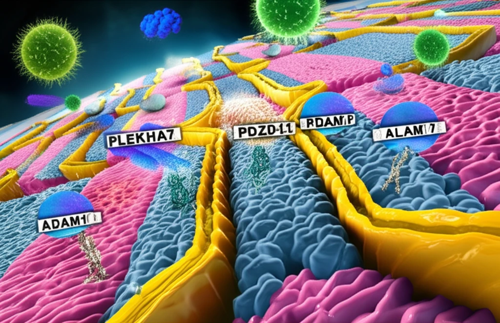
- Docking ADAM10: The transmembrane partner Tspan33 docks ADAM10 to junctions through its cytoplasmic C terminus binding to the WW domain of PLEKHA7 in the presence of PDZD11.
- Locking ADAM10: ADAM10 is locked at junctions through binding of its cytoplasmic C terminus to afadin.
- Efficient Pore Formation: Junctionally clustered ADAM10 supports the efficient formation of stable toxin pores.
- Removal of Toxins: Loss of PLEKHA7-PDZD11 leads to toxin pore removal by endocytosis and cell survival.
Emerging Insights at the Toxin-Junction Interface
Research into how bacterial toxins interact with cellular junctions continues to evolve, though comprehensive reviews on this specific intersection remain limited. The field is moving toward understanding not just how toxins damage cells, but how they co-opt normal cellular machinery to do so. Advances in cellular microbiology are increasingly revealing the intricate molecular dialogues between pathogens and host cell architecture. As investigative tools improve, previously overlooked mechanisms of junction-mediated toxin vulnerability are expected to come to light.
Definitional Boundaries and Target Specificity Debates
Bacterial toxins are defined as harmful substances released by bacteria that cause tissue damage either through direct cellular injury or by provoking an immune response. However, some bacterial toxins recognize ubiquitous membrane components as receptors and indiscriminately damage membranes from different cells, including those of neuronal origin. This raises questions about how specific the "dock-and-lock" mechanism at junctions truly is, versus a more generalized membrane disruption. The tension between targeted junction exploitation and broad membrane damage remains an area of active investigation.
Context-Dependent Toxin Mechanisms Across Pathogen Species
Different bacterial species appear to exploit junctions in distinct ways, suggesting that the dock-and-lock mechanism may be one of several strategies rather than a universal rule. The degree to which a pathogen targets junctions versus other cellular entry points likely depends on the specific tissue tropism and infection niche of each bacterium. As researchers compare mechanisms across species, it becomes increasingly clear that context—in terms of cell type, tissue environment, and bacterial strategy—shapes how junctions factor into toxin delivery. These comparisons underscore the importance of studying multiple pathogen systems rather than generalizing from a single model.
A New Path to Fighting Infections
This discovery of the 'dock-and-lock' mechanism has significant implications for how we approach treating S. aureus infections. Rather than simply targeting the toxin itself, a potential approach involves disrupting the interaction between PLEKHA7 and ADAM10 at cell junctions. By preventing the clustering of ADAM10, cells could be rendered less susceptible to alpha-toxin, giving the body's natural defenses a better chance to clear the infection. Future research will focus on developing drugs that specifically target the PLEKHA7-PDZD11-ADAM10 complex, offering a more targeted and potentially less toxic approach to combating S. aureus and other bacterial pathogens.
Reframing Our Understanding of Cellular Vulnerability
The emerging picture of junction-mediated toxin entry challenges the traditional view of cell junctions as purely protective structures. What were once seen solely as barriers that keep pathogens out may also serve as unintended portals that facilitate toxin entry under certain conditions. This duality—junctions as both shield and potential vulnerability—represents a significant conceptual shift in how researchers think about epithelial defense. As the field matures, integrating junction biology with toxinology will be essential for a complete understanding of host-pathogen interactions.
Targeting Junctions to Combat Infection
A pore-forming toxin produced by Listeria monocytogenes induces changes in the host cell cytoskeleton that facilitate cell-to-cell spread of this intracellular bacterial pathogen, illustrating how toxins can co-opt junctional machinery for dissemination. New research reveals how cell junctions, typically protectors, can become accomplices in bacterial toxin attacks, potentially revolutionizing approaches to fighting infections. If the dock-and-lock mechanism proves tractable as a therapeutic target, it could open doors to interventions that block toxin entry without disrupting the essential barrier functions of junctions. The intersection of junction biology and toxinology represents a promising frontier for next-generation anti-infection strategies.
Scaling From Bench to Bedside
Translating junction-targeting insights from laboratory models to clinical interventions faces significant hurdles, including the complexity of human tissue architecture and the diversity of bacterial strategies. Developing therapies that selectively disrupt toxin-junction interactions without compromising the structural integrity of epithelial barriers will require precision engineering at the molecular level. The broader challenge of antimicrobial resistance further complicates the landscape, as new anti-virulence approaches must prove their value alongside or in combination with existing antibiotics. Ultimately, addressing these systemic challenges will demand interdisciplinary collaboration spanning microbiology, cell biology, and pharmacology.
From Molecular Discovery to Clinical Application
The discipline of cellular microbiology has proven fruitful in shaping our understanding of eukaryote processes through studies of bacterial toxin interference. A recent study has uncovered a dock-and-lock mechanism that controls how ADAM10 is clustered at cell-cell junctions, influencing the cell's vulnerability to alpha-toxin. This mechanism may hold the key to developing new strategies for preventing bacterial infections, bridging the gap between fundamental molecular biology and tangible clinical outcomes. As researchers continue to unravel how pathogens exploit our own cellular machinery, the potential for precision interventions that prevent disease at its earliest molecular steps grows increasingly real.