
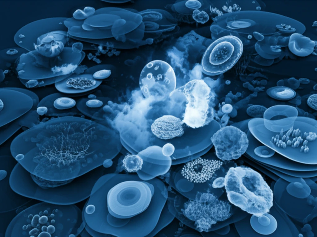
Cell Cultures: Can 3D Models Revolutionize Disease Treatment?

"Discover how 3D cell cultures offer a more realistic way to study diseases and test treatments, potentially changing how we approach medical research and drug development."
In the ever-evolving landscape of medical research, a groundbreaking technique is gaining momentum: three-dimensional (3D) cell cultures. For years, scientists have relied on traditional two-dimensional (2D) cell cultures to study diseases and develop new treatments. However, these 2D models often fall short of replicating the complex environment within the human body.
That's where 3D cell cultures come in. Unlike their 2D counterparts, 3D cell cultures create a more realistic spatial, biochemical, and cellular environment. This allows researchers to study cell behavior, disease progression, and treatment responses in a way that closely mimics what happens in the body.
This article delves into the world of 3D cell cultures, exploring their benefits, applications, and potential to revolutionize disease treatment. From cancer research to drug development, discover how 3D models are paving the way for more effective and personalized medicine.
Why 3D Models Matter
Three-dimensional cell culture is gaining momentum in medicine because it can mimic real tissues and provide more accurate biological data than traditional methods. Current models include organoids, spheroids, and organ-on-a-chip systems, which provide physiologically relevant platforms for drug discovery and disease modeling. Reviews identify standardization and simpler protocols as necessary for wider adoption, while integration with artificial intelligence may further improve the predictive power of preclinical studies.
What Are 3D Cell Cultures and Why Are They Important?
Traditional 2D cell cultures involve growing cells in a single layer on a flat surface. While this method has been valuable for basic research, it fails to capture the intricate interactions between cells and their surroundings within a living organism. The human body is a complex, three-dimensional environment where cells communicate and interact in numerous ways.
- Improved Realism: 3D cultures better mimic the in vivo environment, providing more accurate insights into cell behavior.
- Enhanced Cell Interactions: Cells in 3D cultures can interact with each other in a more natural way, influencing growth, differentiation, and response to stimuli.
- Better Drug Testing: 3D models can provide a more reliable platform for testing new drugs, as they can predict treatment responses more accurately than 2D cultures.
- Personalized Medicine: 3D cultures can be created using a patient's own cells, allowing for personalized testing of treatments and therapies.
New Directions in 3D Research
The transition from two-dimensional to three-dimensional models is described as a pivotal advancement in biomedical research. Spheroids, organoids, and organ-on-a-chip systems offer more physiologically relevant environments that can improve predictions of drug efficacy, toxicity, and disease mechanisms. Recent reviews also highlight tumor spheroids, tumor organoids, and tumor-on-chip models for cancer research, metastasis and invasion studies, personalized therapeutics, and drug screening.
Persistent Limitations
Although 3D models show reported benefits over conventional 2D cultures, reviews continue to examine their limitations, applications, and preclinical testing methods. Traditional 2D cultures remain widely used, especially in cancer biology, but their limitations have motivated the development of three-dimensional systems. The reported advantages of 3D culture include better representation of tissue structure and function, improved cell morphology and interactions, and potentially greater predictability in drug research.
Comparing 2D and 3D Models
Comparative studies have examined standard 2D tissue-culture plates alongside classical 3D systems using commercially available Engelbreth-Holm-Swarm tumor-derived gel and synthetic scaffolds. A December 2024 study specifically compared 2D and 3D models as preclinical systems for responses to carboplatin, paclitaxel, and niraparib. That work used PEO1, PEO4, and PEO6 cell lines generated from the same patient during disease progression, illustrating how model choice can be evaluated in a defined treatment context.
The Future of Disease Treatment with 3D Cell Cultures
3D cell cultures represent a significant step forward in medical research, offering a more realistic and reliable way to study diseases and test new treatments. While 2D cell cultures have served as a valuable tool for decades, they often fail to capture the complexity of the human body. 3D models, on the other hand, provide a more accurate representation of cell behavior and disease progression, paving the way for personalized medicine and more effective therapies. As technology continues to advance, the potential of 3D cell cultures to revolutionize disease treatment is only just beginning to be realized, and this is what to keep an eye on.
Toward Better Disease Models
A 2021 review reports that co-culturing cells in 3D offers advantages over 2D cell culture. It connects advances in tissue-engineering techniques with expected improvements in tumor models, cancer treatments, and disease-testing methods. The review therefore presents 3D culture as part of a broader progression toward more advanced biomedical modeling.
The Road Ahead
Future development is expected to focus on 3D systems that mimic the natural cellular environment while becoming easier to standardize and use. A 2026 Special Issue describes new models across broad biomedical areas and emphasizes their potential contributions to targeted therapies and biomedical engineering. The biopharmaceutical industry is also reported to be showing increased demand for cell-culture products that support drug discovery and safety testing.
From Models to Medicine
Reported case studies describe real-world applications of 3D cell culture in cancer research, drug discovery and testing, and tissue engineering and regenerative medicine. These examples frame 3D culture not only as a laboratory technique but also as a tool connected to treatment development and tissue repair. The available source identifies these areas of impact without providing specific study names or outcome figures.