
Cam Hip Morphology: Are You at Risk?

"Unveiling the prevalence and potential implications of cam hip morphology in the general population."
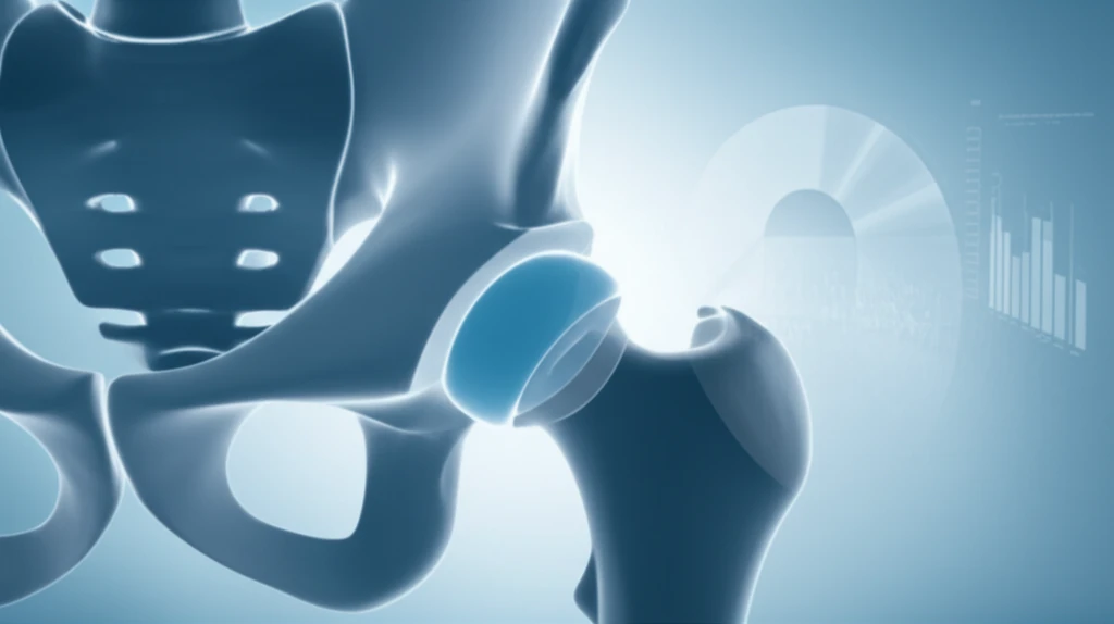
Femoroacetabular impingement (FAI) syndrome, a motion-related hip disorder, arises from premature contact between the femur and acetabulum. This condition often stems from specific hip shapes, including cam and pincer morphologies. Cam morphology is characterized by a flattening or convexity at the femoral head-neck junction, leading to impingement against the acetabular rim during movement.
The presence of cam morphology has long been linked to the development of hip osteoarthritis (OA). Early observations in the 1960s noted a high prevalence of cam hip shapes in patients undergoing hip arthroplasty. Later research identified FAI as a potential cause of hip OA, with studies demonstrating an increased OA risk in individuals with cam morphology.
Despite growing recognition of cam morphology's role in hip OA, its precise prevalence remains unclear. Initial systematic reviews revealed prevalence estimates ranging widely, from 5% to 75% of the general population. The lack of uniform diagnostic measures and diverse study populations hindered meta-analysis and definitive conclusions.
Numbers That Matter: The World COACH Evidence
Tang, J. et al.'s individual participant data meta-analysis of 23,886 hips from the Worldwide Collaboration on Osteoarthritis PrediCtion for the Hip (World COACH) consortium assessed whether cam morphology predicts radiographic hip osteoarthritis (RHOA) developing within 8 years, selecting hips free of RHOA at baseline with 4–8 years of follow-up. The analysis examined the relationship overall and in subgroups based on age, biological sex, and body mass index (BMI). Complementary prospective work tracks the same question in smaller cohorts: a nationwide CHECK cohort study with 10 years of follow-up uses statistical shape modeling to define cam subtypes and their relationship with hip OA, while an Osteoarthritis and Cartilage analysis followed up at four time points across a 10-year window.
How the Field Measures Cam Morphology
Across the literature reviewed here, the standard approach is observational: identify cam morphology on imaging, typically radiographs, and then follow participants forward over several years to see whether radiographic hip osteoarthritis develops. Cam morphology is generally defined by a bony prominence at the femoral head–neck junction, but the specific definitions and measurement thresholds used can vary from study to study. These methods carry recognized limitations, including the possibility that a single imaging view misses subtle or borderline morphology and that different classification schemes make results hard to compare directly. Readers should therefore treat the reported associations as current research findings that may shift as measurement approaches evolve.
From Shape Variation to Risk Factor
Cam morphology is formally defined as a bony prominence around the femoral head–neck junction of the hip that alters the normal shape of the femoral head, which makes it one of the most common hip morphologies and an important factor in the development of hip osteoarthritis. A Review published in November 2025 describes how this shape variation can cause intra-articular joint damage and lead to femoroacetabular impingement (FAI) syndrome and, eventually, hip osteoarthritis. The same Review emphasizes that early detection of the related FAI syndrome is crucial for timely intervention and for potentially preventing progression to osteoarthritis.
Understanding Cam Morphology: Prevalence and Impact
A recent study aimed to define the prevalence of cam morphology within the general population using cross-sectional imaging and a predefined diagnostic accuracy measure. The research involved screening patients presenting to University Hospitals of Coventry and Warwickshire (UHCW) after major trauma.
- 47% of subjects aged 16-65 had cam morphology.
- 56% of males and 37% of females were affected.
- The sample was broadly representative of the UK general population.
- Cam morphology was measured using cross sectional imaging.
What Recent Reviews Conclude
Recent reviews continue to describe cam morphology as one of the most common hip morphologies—a bony prominence around the femoral head–neck junction that alters the shape of the femoral head—and a key risk factor for hip osteoarthritis. A 2025 review (PubMed 41298958) highlights that early detection of the related femoroacetabular impingement (FAI) syndrome is crucial for timely intervention and potentially for preventing osteoarthritis progression. Complementing that, a 2023 Osteoarthritis and Cartilage analysis found that cam morphology is strongly and consistently associated with the development of radiographic hip osteoarthritis at four follow-up visits within a 10-year window, with FAI proposed as a mechanism for the development of early osteoarthritis.
Caveats and Open Questions
The evidence linking cam morphology to later hip osteoarthritis is substantial, but the research is not without caveats. Not every hip with cam morphology will go on to develop osteoarthritis, and the material reviewed here does not establish a causal chain or a precise level of individual risk. Findings are sensitive to how cam morphology is defined, measured, and followed over time, which means results can differ across studies. For these reasons, cam morphology is best characterized as a risk factor rather than a guaranteed predictor of joint disease.
Comparing Designs, Durations, and Populations
Because the research in this area varies in design—from large meta-analyses pooling many hips to single prospective cohorts—findings are not always directly comparable across studies. Follow-up windows, population types, imaging methods, and classification schemes can all differ, which limits how confidently any single risk figure can be generalized. The safest reading of the literature is that the direction of the association between cam morphology and hip osteoarthritis is consistent, even though the exact magnitude remains unsettled. Any comparison of specific numbers from different studies should therefore be made with caution.
What Does This Mean For You?
This study highlights the relatively high prevalence of cam morphology in the general population. Although the presence of cam morphology does not automatically equate to FAI syndrome, it may increase the risk of developing hip osteoarthritis.
The Consistent Thread
Taken together, the research points to a single central conclusion: cam morphology is consistently linked with an increased likelihood of developing hip osteoarthritis, particularly when identified on radiographs and tracked over several years. The supporting evidence spans everything from a large international meta-analysis of nearly 24,000 hips to smaller prospective cohort studies with follow-up as long as a decade. However, because the exact strength of the association and the most reliable detection method remain active questions, interpretation should be appropriately cautious. The practical implication for readers is to regard cam morphology as a notable risk factor worth discussing with a clinician, not as a diagnosis of existing joint disease.
X-Rays as a Screening Signal
A January 2026 BJSM blog post reporting on the World COACH meta-analysis offers a clear near-term outlook: cam morphology, as identified on standard anteroposterior (AP) pelvic radiographs, is a significant and independent risk factor for developing hip osteoarthritis within 4–8 years. The post describes the study as providing robust evidence, presenting key take-home points that plain radiographs could serve as a practical first-line signal for flagging at-risk hips. This points toward risk stratification in routine clinical settings rather than immediate diagnosis or treatment. Future research will likely continue testing how reliably radiograph-based screening translates into earlier attention and, ultimately, prevention.
Population-Level Gaps
A prospective cohort study that analyzed anteroposterior hip radiographs from 1,019 participants notes that cam morphology is understood to contribute to the development of hip osteoarthritis but has been less studied in the general population, and it flags limits to generalizing findings to the broader population. The related Osteoarthritis and Cartilage research stream set out to determine the association between cam morphology and the development of radiographic hip osteoarthritis at four time points within a 10-year follow-up. Together these sources illustrate a systemic challenge: much of the evidence base comes from defined research cohorts, so population-wide estimates of how commonly cam morphology advances to osteoarthritis remain uncertain. Closing that gap will require larger, more representative samples and more consistent definitions across settings.
What This Means for One Person
Cam morphology is a subclinical shape variation, present in hips that may never cause symptoms, which is precisely why the research findings matter personally. For someone told they have cam morphology, the realistic concern is not necessarily immediate pain but the possibility of femoroacetabular impingement symptoms and, over years, an elevated chance of hip osteoarthritis that could affect mobility and daily activity. Because detection is often incidental—an X-ray taken for another reason—the practical takeaway is a conversation with a clinician about symptoms, activity, and whether monitoring is warranted, rather than alarm. The research cannot yet tell any single individual their precise risk, but it gives patients and doctors a reason to take the finding seriously and plan ahead.
If you experience hip pain, especially during movement, consult with a healthcare professional. Early diagnosis and management can help mitigate potential complications associated with FAI syndrome and cam morphology.
Future research should focus on prospective studies assessing the association between hip pain, clinical findings, and hip morphology to better understand the epidemiology of FAI syndrome and guide preventative strategies.