
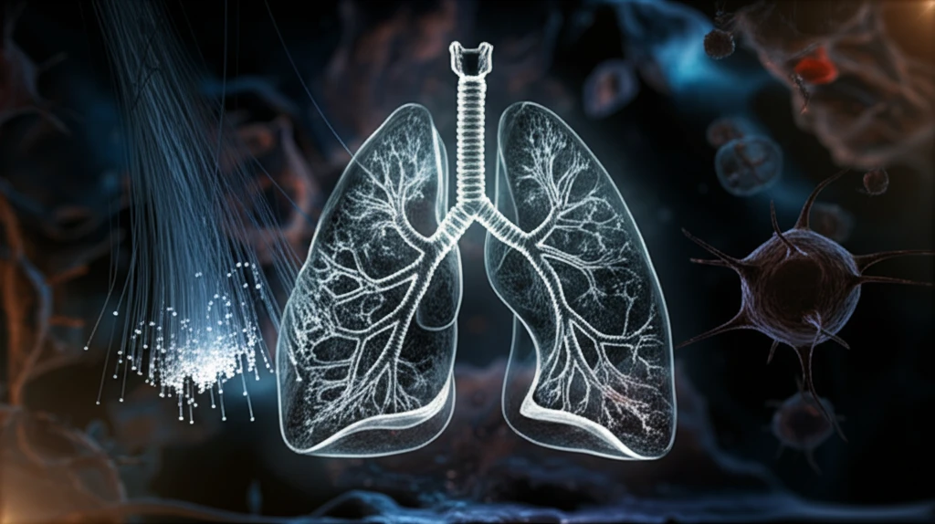
Breathe Easier: The Latest Breakthroughs in Early Lung Cancer Detection

"Combining Fiberoptic Bronchoscopy and Liquid-Based Cytology for a More Accurate Diagnosis"
Lung cancer remains a significant global health challenge, with increasing incidence and mortality rates. Early detection is crucial for improving survival rates, as treatment is far more effective in the early stages of the disease. While advancements in treatment have been made, the key to combating lung cancer lies in identifying it as early as possible.
Traditional methods for diagnosing lung cancer often struggle to detect early-stage tumors due to their small size or location. Fiberoptic bronchoscopy allows doctors to visualize the airways, but it may miss subtle signs of cancer. Cytology, the study of cells, can help, but traditional methods of collecting and analyzing cell samples have limitations.
Now, a promising new approach combines fiberoptic bronchoscopy with liquid-based cytology (LCT) to enhance early detection. This technique aims to overcome the limitations of previous methods, offering a more accurate and reliable way to diagnose lung cancer in its earliest, most treatable stages.
Early Detection Saves Lives
Low-dose CT (LDCT) scanning is a quick, painless, and noninvasive exam that takes detailed images of the lungs, detecting small spots or nodules long before symptoms appear, when treatment is most effective. Early detection can be life-saving because it finds cancer while it is still confined to the lung and has not spread to lymph nodes or distant organs, typically classified as Stage I. Such early findings give patients and doctors the best window for successful intervention.
Standard Tools: From LDCT Scans to Bronchoscopy
Low-dose CT screening is the accepted standard method for early detection, offering a fast, painless exam that has been proven to save lives. Yet many basic health screening packages may miss early lung changes, which is why dedicated screening programs matter. When follow-up is needed, bronchoscopy—an endoscopic technique for visualizing the inside of the airways—can be used for diagnostic and therapeutic purposes, and a fiberoptic bronchoscopy allows doctors to examine the airways, take biopsy samples, and perform minor treatments.
A Tradition of Airway Visualization
Bronchoscopy has long served as a foundational diagnostic tool, with the technique of visualizing the inside of the airways evolving over time for both diagnostic and therapeutic purposes. As imaging technology advanced, low-dose CT scanning emerged as a noninvasive screening method that can image the lungs without an instrument inserted into the airways. These complementary approaches represent the milestones that shaped modern lung cancer detection.
The Power of Combined Techniques
A recent study published in Oncology Letters investigated the effectiveness of combining fiberoptic bronchoscopy with liquid-based cytology (LCT) in the early diagnosis of lung cancer. The study retrospectively analyzed data from 901 patients with suspicious lung lesions who underwent bronchoscopy with LCT at Shanxi Provincial Cancer Hospital between June 2012 and June 2016.
- Group A: Bronchoscopic washing cultures (BWC)
- Group B: Bronchoalveolar lavage (BAL)
- Group C: Endobronchial ultrasound-guided transbronchial needle aspiration (EBUS-TBNA)
- Group D: Transbronchial lung biopsy (TBLB)
A Breath of Fresh Air: New Testing Frontiers
Recent research has explored a new breath test breakthrough that could aid in early lung cancer detection, using a sensor that can measure isoprene in human breath with remarkable precision. In the study, isoprene levels were found to be lower in lung cancer patients compared to those without cancer. Researchers also note that lung cancer may present differently in women, highlighting the need for detection approaches that account for varied symptoms and disease patterns.
Why General Screenings Can Fall Short
Not all screening approaches are equally effective at catching early lung cancer. Many basic health screening packages may miss early lung changes, underscoring why specialized low-dose CT screening is recommended over generic check-ups. Because the disease can also present differently in women, a one-size-fits-all approach may overlook cases that do not follow typical patterns, making awareness of varied presentations an important part of early detection.
Comparing Detection Strategies
Low-dose CT screening provides a fast, painless, and noninvasive way to image the lungs and spot nodules before symptoms develop, whereas basic health screening packages may not be sensitive enough to catch early lung changes. For patients who need deeper investigation, bronchoscopy offers direct visualization of the airways for diagnostic purposes, including biopsy sampling. Each method serves a distinct role: CT for population-level early detection, and bronchoscopy for confirming and characterizing suspected findings.
A Brighter Future for Lung Cancer Diagnosis
The study's findings suggest that fiberoptic bronchoscopy combined with liquid-based cytology is a safe, reliable, and effective technique for the early diagnosis of lung cancer. By improving the accuracy of early detection, this approach offers the potential to significantly improve treatment outcomes and survival rates for individuals at risk of lung cancer.
Experts Agree: Stage I Is the Goal
Experts emphasize that catching lung cancer early can be life-saving. When lung cancer is found 'early,' it means the cancer is confined to the lung and has not spread to lymph nodes or distant organs, typically classified as Stage I. Screening programs describe LDCT as a proven tool that saves lives, and patient stories reinforce that an early catch can make a critical difference.
Breath-Based Diagnostics on the Horizon
Breath-based diagnostics represent a promising frontier in early lung cancer detection. A newly reported sensor can measure isoprene in human breath with remarkable precision, and study results show isoprene levels are lower in lung cancer patients than in those without cancer. If refined, such a breath test could offer a fast, noninvasive complement or alternative to imaging-based screening.
Access and Eligibility Remain Hurdles
Widespread early detection depends on access to screening, and many providers promote free screening locations and eligibility checks, such as those offered around World Lung Day. Patients are encouraged to use location searches to find nearby facilities and to confirm whether they qualify for low-dose CT screening. Health experts frame screening not merely as a check of whether you feel healthy today, but as an investment in future health.
Real Stories: Catching Cancer Early
Personal stories illustrate the real-world impact of early detection. Beth Mathis had her cancer caught so early that, in her words, it was 'caught before it even had a chance to grow,' and a robotic surgery to remove the lower lobe of her right lung left her in the hospital for just two days. Public figures like Barry Manilow, at 82, have also shared health updates on lung cancer, underscoring that early diagnosis matters at every stage of life.