
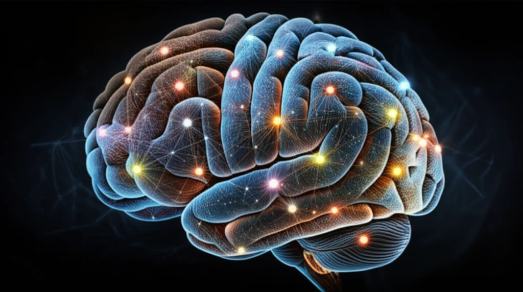
Brain Graphs: Are We Mapping Our Minds the Right Way?

"A new study reveals that how we define the nodes in brain network analysis significantly impacts the accuracy of our mental maps."
The human brain, a vast and intricate landscape of interconnected regions, has always been a subject of fascination. Modern neuroscience uses functional Magnetic Resonance Imaging (fMRI) to peek inside this landscape, mapping the complex communication networks that orchestrate our thoughts, emotions, and actions. Graph theory, a mathematical framework for studying networks, has become a popular tool for analyzing these brain networks, offering a way to quantify their architecture and understand how they function in both health and disease.
But there's a fundamental challenge in constructing these brain graphs: How do we define the 'nodes,' the basic units of the network? Two primary methods dominate the field. The first involves using predefined regions of interest (ROIs) based on brain atlases. Imagine dividing the brain into a map of territories, each representing a specific anatomical area. The second approach, independent component analysis (ICA), is data-driven. It identifies independent components or spatial brain maps directly from the fMRI data, letting the brain's activity dictate the network's structure.
The question then becomes: Which method provides a more accurate representation of the brain's true functional organization? It's a debate that has significant implications for how we interpret brain network studies. A groundbreaking study steps into this arena, using simulated fMRI data to compare the performance of brain graphs constructed with ROI-based nodes versus ICA-based nodes. The findings challenge conventional wisdom and point towards a potentially more effective way to map our minds.
The Brain’s Central Role
The brain is the center of the nervous system in vertebrate and most invertebrate animals, receiving information from the sensory nervous system. In humans, it processes, integrates, and coordinates sensory information, then directs instructions to the rest of the body; the cerebrum is its largest part and has two cerebral hemispheres. Johns Hopkins Medicine also describes the brain as controlling thought, memory, emotion, touch, motor skills, vision, respiration, and other processes that regulate the body.
A Complex Coordinated System
The brain regulates movement, memory, emotions, thoughts, body temperature, breathing, hunger, and many other aspects of daily functioning. Cleveland Clinic emphasizes that many complex parts work together to support these functions. This complexity means that a broad description of the brain’s roles does not, by itself, explain how its parts interact or how those interactions produce behavior.
An Evolving Map
The history of brain research can be understood as a gradual effort to connect physical structures with sensation, movement, thought, and behavior. Earlier models tended to simplify the brain into separate parts, while later approaches increasingly recognized interactions across regions. This broad shift suggests that brain mapping has developed through successive approximations rather than a single final account.
ICA vs. ROI: Unpacking the Brain Mapping Methods
To understand the study's results, let's delve deeper into the two competing methods for defining nodes in brain graphs:
- Pros: Straightforward, easy to implement, and provides a standardized way to compare results across studies.
- Cons: ROIs are based on anatomical boundaries, which may not perfectly align with functional boundaries. This means that an ROI might contain a mix of functionally distinct areas, blurring the picture of brain activity.
Research Beyond Simple Maps
Current brain research is increasingly likely to treat the brain as a dynamic system rather than a fixed collection of isolated locations. Researchers may combine structural, functional, and behavioral evidence to examine how activity changes across networks. Because this area remains active and developing, individual findings should be interpreted cautiously until they are replicated and connected to broader evidence.
The Limits of Simplification
A brain graph can make complex relationships easier to visualize, but a visual network is not necessarily a complete explanation of cognition. Connections may correlate with a mental process without uniquely causing it, and the same behavior may involve multiple interacting systems. These limitations mean that attractive maps should not automatically be treated as definitive accounts of how the mind works.
Comparing Models
Different approaches to understanding the brain emphasize different kinds of evidence, including anatomy, activity, connectivity, and behavior. A region-based model may be easier to interpret, while a network-based model can better represent interactions that cross traditional boundaries. No single comparison can establish that one model is universally best, because usefulness depends on the question being asked and the evidence available.
The Future of Brain Mapping
This study offers compelling evidence that the choice of node definition significantly impacts the accuracy of brain graph analysis. By demonstrating the superior performance of ICA-based methods in simulated fMRI data, the researchers challenge the conventional reliance on ROI-based approaches. The implications are far-reaching, suggesting that data-driven methods may hold the key to unlocking deeper insights into brain function and dysfunction.
Maps as Working Models
Brain graphs are best treated as working models that organize evidence rather than as literal pictures of the mind. Their value lies in linking patterns of neural organization with functions while preserving uncertainty about causation and interpretation. A careful synthesis therefore combines network descriptions with behavioral, clinical, and biological context.
Toward More Complete Models
Future brain mapping may focus on models that connect changing neural activity with behavior over time. Progress will likely depend on integrating multiple measurement methods and testing whether observed network patterns generalize across people and situations. The next frontier is therefore not simply drawing larger graphs, but determining which connections are meaningful, reproducible, and useful.
The Challenge of Scale
Brain mapping operates across several scales, from microscopic connections to whole-brain systems and observable behavior. Aligning these scales is difficult because a pattern visible at one level may not directly explain what happens at another. Broader interpretation also requires attention to uncertainty, measurement limits, and differences among individuals.
From Networks to People
The significance of brain graphs ultimately depends on whether they improve understanding of real human experience. A map may support research or clinical reasoning, but it cannot replace attention to a person’s symptoms, history, environment, and goals. Keeping that human context visible can help prevent technical representations from being mistaken for complete accounts of individual lives.