
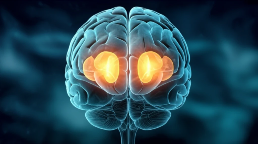
Illustration of an adolescent brain highlighting asymmetric subcortical regions.

Brain Asymmetry and Mental Health: What Early Brain Scans Reveal About Psychotic Experiences

"New research uncovers how subtle differences in brain structure during adolescence might offer clues to future mental health challenges, sparking new avenues for early intervention and support."


Mental health disorders, particularly those involving psychosis, are complex and often devastating. Scientists have long sought to understand the biological underpinnings of these conditions, hoping to identify early markers that could allow for timely intervention. A recent study published in 'Translational Psychiatry' sheds new light on this area by exploring the relationship between brain structure and subclinical psychotic experiences (SPEs) in adolescents.

SPEs are experiences that resemble psychotic symptoms, such as hallucinations or delusions, but are less severe and don't meet the criteria for a full-blown psychotic disorder. These experiences are not uncommon in adolescence, with some studies estimating that up to 5% of young people may experience them. While many individuals who report SPEs do not go on to develop psychosis, they are at an increased risk, making the identification of potential biological markers crucial.

The study, led by researchers at the University of Tokyo, investigated whether differences in the volumes of subcortical brain regions are associated with SPEs in a large sample of adolescents. Subcortical structures, including the basal ganglia and limbic system, play critical roles in various functions, including learning, emotion, motor control, and higher-order cognitive processes. Previous research has suggested that alterations in these regions may be linked to schizophrenia and other psychotic disorders.

AI Search Multiple angles on this topic

Volumetric and VBM Measures

The cited study concerns volumetric and voxel-based morphometry (VBM) measures in children aged 1.5 to 5 years obtained using adult segmentation tools. Its source material places psychosis, including Okada et al. (2018), among the conditions discussed in relation to these neuroimaging measures. The snippet also mentions epilepsy and anxiety, but it does not provide prevalence figures or quantified mental-health impact.

Measuring Brain Asymmetry

Brain asymmetry is studied through functional and structural measures, with the asymmetry index (AI), also called the laterality index, commonly used to quantify cerebral asymmetry. Laterality refers to the dominance of one hemisphere in particular activities or functions, including handedness and language processing. However, methodological choices vary considerably across neuroimaging techniques and research contexts. Recent work also emphasizes that asymmetry develops dynamically and is associated with individual variability, cognitive outcomes, and psychiatric outcomes during development.

Unveiling the Adolescent Brain: What the Study Showed

Illustration of an adolescent brain highlighting asymmetric subcortical regions.

The researchers used magnetic resonance imaging (MRI) to measure the volumes of subcortical brain structures in 203 adolescents aged 10.5 to 13.3 years. These participants were part of a larger population-based cohort study called the Tokyo TEEN Cohort (TTC). The adolescents also completed a screening questionnaire to assess their experiences with SPEs.

The study's findings revealed several significant associations between brain volume and SPEs. Adolescents with SPEs had larger volumes in the left hippocampus, right caudate, and right lateral ventricle compared to those without SPEs. They also found a marginally significant increase in the left pallidum.

  • Left Hippocampus: This brain area is vital for memory and learning.
  • Right Caudate: Part of the basal ganglia, involved in motor control and cognitive functions.
  • Right Lateral Ventricle: A fluid-filled space in the brain; increased volume may indicate various neurological conditions.
  • Left Pallidum: Another component of the basal ganglia, important for motor control.
AI Search Multiple angles on this topic

Regional and Subcortical Findings

Recent research links negative symptoms in schizophrenia patients with altered structural asymmetry in the amygdala and superior temporal region. Previous studies reported rightward amygdala-volume asymmetry in both healthy controls and schizophrenia patients, but did not find significant differences between those groups or between adolescents with and without subclinical psychotic experiences. A mega-analysis of 5,604 subjects reported lateralization findings involving pallidum volume in early-onset psychosis, at-risk mental states, and antipsychotic-naive adolescents with subthreshold psychotic experiences. Reviews also describe widespread structural and functional differences across frontal, temporal, and parietal lobes and the basal ganglia, while one study found no significant regional-volume difference between at-risk groups with and without later psychosis onset.

Limits of Early Evidence

Evidence for asymmetry as a premorbid marker remains tentative. One study proposed that leftward alteration of pallidal-volume laterality could be possible premorbid neuropathology in schizophrenia, but also noted methodological limitations. In particular, the Adolescent Psychotic Symptoms Survey data were collected on average 16 months before MRI scanning, so the reliability of the results could not be fully guaranteed. Broader reviews describe atypical hemispheric asymmetries as a possible mental-health consequence while emphasizing that the field is still developing.

Developmental Timing and Disorder

Cerebral lateralization is described as a core organizing principle involving hemispheric specialization and interactions between the hemispheres. Across mental disorders, functional and structural hemispheric asymmetries may differ from those observed in healthy controls, and these alterations may contribute to symptoms and cognitive impairments. A key comparative question is the temporal sequence of asymmetry formation relative to early pathophysiology. Determining when asymmetrical differentiation occurs in disorder-relevant brain regions is necessary for evaluating possible developmental relationships.

Perhaps most interestingly, the researchers found that adolescents with SPEs showed significantly more leftward laterality of pallidal volume than those without SPEs. This means that the left pallidum was larger than the right pallidum in individuals with SPEs, a pattern that mirrors findings in adults with chronic schizophrenia. This discovery suggests that altered pallidal asymmetry may be a premorbid characteristic, potentially indicating a predisposition to developing psychosis later in life.

What Does This Mean for the Future of Mental Health?

This research offers valuable insights into the early neurobiological underpinnings of psychotic experiences. By identifying potential markers like altered pallidal asymmetry, scientists may be able to develop more effective strategies for early identification and intervention. Further research is needed to fully understand the role of these brain differences and how they may interact with genetic and environmental factors. However, this study represents an important step forward in our quest to unravel the complexities of mental illness and improve the lives of those at risk.

AI Search Multiple angles on this topic

From Localized to Global Asymmetry

Future research is being directed toward a multivariate view of global brain asymmetry. Traditional work has often focused on localized interhemispheric differences between homologous pairs of regions. The proposed integrative perspective instead examines hemispheric differences as global patterns across the entire brain. This approach opens new questions about how the hemispheres are organized and provides a direction for further investigation.

How adolescent pallidal findings compare with later reports

Later papers cited this leftward pallidal pattern in adolescents alongside reports involving at-risk mental states and early-onset psychosis, while noting that lateralization in early-onset psychosis had not been investigated in detail.[1]

A later schizophrenia analysis described increased leftward pallidal asymmetry in schizophrenia as also detectable in adolescents with SPEs, placing the adolescent observation within a broader comparison across groups.[2]

Structural MRI and subcortical volumes in early adolescents

Study designObservational study[3]
Sample size203[3]
SettingLarge-scale population-based cohort[3]
Effect sizeCohen’s d effect sizes and standard errors were calculated for group differences in subcortical regional volumes[3]
p-valuep = 0.022–0.023[3]
Times cited45[3]

What a cross-sectional cohort study can establish

Observational study (level 4 of 8)

Because the study was observational and cross-sectional, it can identify an association between SPEs and brain measurements at the time studied, but cannot establish that either caused the other.[3] [4]

The population-based cohort context gives the findings relevance to adolescents beyond a clinical patient sample, but the measured subsample remains limited in size and age range.[3]

The pallidal comparison depends on the subgroup

  • 1Among adolescents limited to Tanner stage 1, those with SPEs had larger left pallidal volume and more leftward asymmetry; this subgroup result sits alongside the overall finding of no significant bilateral pallidal-volume differences.[4]Caveat: Overall bilateral pallidal-volume differences were not significant.

What the brain differences do and do not imply

The authors frame leftward pallidal asymmetry as a possible feature related to later schizophrenia, not proof that an adolescent with SPEs will develop the disorder; the study did not test causation.[3] [4]

Other regional differences were interpreted cautiously: the authors described increased left hippocampal volume as possibly compensatory, while the reason for increased right caudate volume remained unclear.[4]

What keeps the pallidal interpretation tentative

  • The authors characterize premorbid pallidal laterality as a possibility, rather than an established marker.[4]

What remains unresolved about asymmetry and risk

  • Do leftward pallidal asymmetries in early adolescence reflect a predisposition to later psychosis, or another feature associated with SPEs?[3] [5]
  • How should a leftward asymmetry be interpreted against the broader population pattern, in which pallidal volume is also larger on the left on average?[3] [6]
  • How do subcortical measurements relate to the dynamic brain maturation described in young people at risk for psychosis?[7]
Laterality index
A ratio comparing left-minus-right volume with the sum of left and right volume, used to assess structural asymmetry.[3]

About this Article -

Written with AI assistance from published research, and reviewed by the Mystum team. See our About page for more information.

This article is based on research published under:

DOI-LINK: 10.1038/s41398-018-0312-6, Alternate LINK

Title: Abnormal Asymmetries In Subcortical Brain Volume In Early Adolescents With Subclinical Psychotic Experiences

Subject: Biological Psychiatry

Journal: Translational Psychiatry

Publisher: Springer Science and Business Media LLC

Authors: Naohiro Okada, Noriaki Yahata, Daisuke Koshiyama, Kentaro Morita, Kingo Sawada, Sho Kanata, Shinya Fujikawa, Noriko Sugimoto, Rie Toriyama, Mio Masaoka, Shinsuke Koike, Tsuyoshi Araki, Yukiko Kano, Kaori Endo, Syudo Yamasaki, Shuntaro Ando, Atsushi Nishida, Mariko Hiraiwa-Hasegawa, Kiyoto Kasai

Published: 2018-11-28

Everything You Need To Know

1

What are Subclinical Psychotic Experiences (SPEs), and why are they important to study?

Subclinical Psychotic Experiences (SPEs) are experiences that resemble psychotic symptoms like hallucinations or delusions, but are less severe and don't meet the criteria for a full-blown psychotic disorder. SPEs are significant because, while many adolescents who experience them do not develop psychosis, they are at an increased risk. Identifying biological markers associated with SPEs could allow for timely interventions.

2

How did the study measure brain structure, and what specific brain regions were examined?

The study used magnetic resonance imaging (MRI) to measure the volumes of subcortical brain structures in adolescents. The specific subcortical structures examined were the left hippocampus, right caudate, right lateral ventricle, and left pallidum. These structures were chosen because they play critical roles in functions like learning, emotion, motor control, and higher-order cognitive processes, and previous research has linked alterations in these regions to schizophrenia and other psychotic disorders.

3

What did the study discover about the brains of adolescents who experience Subclinical Psychotic Experiences (SPEs)?

The study found that adolescents with Subclinical Psychotic Experiences (SPEs) had larger volumes in the left hippocampus, right caudate, and right lateral ventricle compared to those without SPEs. A marginally significant increase was also found in the left pallidum. Most notably, adolescents with SPEs showed more leftward laterality of pallidal volume, meaning the left pallidum was larger than the right. This finding is significant because altered pallidal asymmetry has also been observed in adults with chronic schizophrenia, suggesting it may be a premorbid characteristic indicating a predisposition to developing psychosis.

4

What are the hippocampus, caudate, lateral ventricle, and pallidum, and what do they do?

The hippocampus, caudate, lateral ventricle, and pallidum are all subcortical brain structures. The left hippocampus is crucial for memory and learning. The right caudate, part of the basal ganglia, is involved in motor control and cognitive functions. The right lateral ventricle is a fluid-filled space; increased volume may indicate various neurological conditions. The left pallidum, another component of the basal ganglia, is important for motor control. Variations in the size or asymmetry of these structures may be linked to mental health conditions.

5

Why is altered pallidal asymmetry considered a potentially important finding?

Altered pallidal asymmetry, where the left pallidum is larger than the right, is significant because this pattern has been observed in both adolescents with Subclinical Psychotic Experiences (SPEs) and adults with chronic schizophrenia. This suggests that altered pallidal asymmetry may be a premorbid characteristic, potentially indicating a predisposition to developing psychosis later in life. Identifying such markers early could allow for interventions to mitigate the risk of developing full-blown psychotic disorders.

Newsletter Subscribe

Subscribe to get the latest articles and insights directly in your inbox.