
Beyond the Image: How Ultrasound Molecular Imaging is Revolutionizing Healthcare

"Discover how contrast agents and cutting-edge techniques are enhancing diagnostics and paving the way for targeted therapies"
Ultrasound molecular imaging represents a significant leap forward in medical diagnostics, offering a non-invasive and highly precise method to visualize biological processes at the cellular level. By integrating ultrasound technology with molecular imaging, clinicians can now detect diseases earlier and with greater accuracy, leading to more effective and personalized treatments. This innovative approach combines the strengths of ultrasound medicine, molecular imaging, materials science, and nanotechnology.
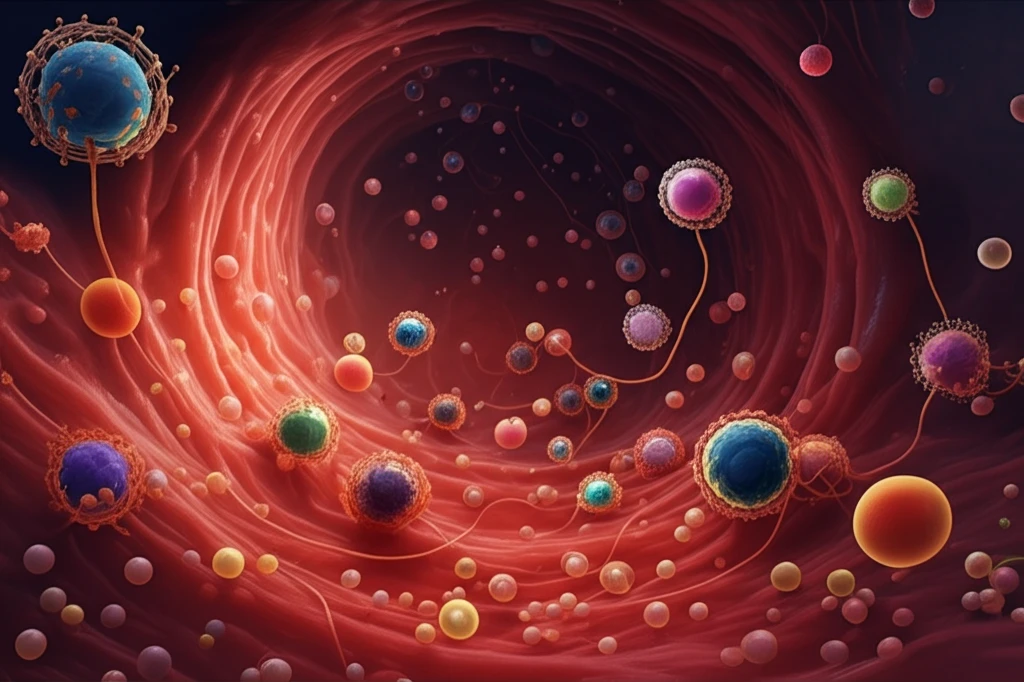
At the heart of ultrasound molecular imaging are ultrasound contrast agents (UCAs), specialized substances designed to enhance the visibility of structures within the body. Traditional ultrasound imaging relies on the reflection of sound waves to create images, but UCAs amplify these signals, providing clearer and more detailed visualizations. These agents, often in the form of gas-filled microbubbles (MBs), are introduced into the bloodstream, where they respond to ultrasound waves, generating enhanced echoes that improve image quality.
The development of nanoscale UCAs has further expanded the capabilities of ultrasound molecular imaging. These tiny agents can penetrate tissues more effectively, allowing for the detection of extravascular lesions and providing new insights into disease processes. By attaching specific ligands to the surface of UCAs, researchers can target them to specific biomarkers, enabling the visualization of tumors, inflammation, and other critical conditions at their earliest stages.
The Growing Impact of Molecular Ultrasound Imaging
Molecular ultrasound imaging is an emerging strategy that combines ultrasound technology with molecularly-targeted contrast agents to assess biological processes at the molecular level. The approach enhances the sensitivity and specificity of ultrasound, allowing visualization of angiogenesis, inflammation, and thrombosis in pre-clinical studies. Contrast agents, particularly microbubbles, are used to amplify the ultrasound signal and provide molecular information. This technology has expanded the diagnostic applications of ultrasound, enabling the evaluation of microvascular blood flow and molecular markers.
Methods, Advances, and Limitations in Molecular Ultrasound
The standard approach involves using targeted contrast-enhanced ultrasound with microbubble contrast agents. While several limitations have historically slowed the progression of ultrasonic molecular imaging, recent advances in contrast agent development, ultrasound technology, and detection strategies show substantial potential. For over 25 years, the scientific understanding of microbubble behavior in vivo and the development of imaging technology have evolved to deliver contrast-enhanced ultrasound. These methods have broadened the scope of medical ultrasound imaging, enabling the assessment and quantification of microvascular blood flow.
Three Decades of Ultrasound Contrast Agents
The diagnostic applications of ultrasound imaging have expanded enormously over the last few decades with the introduction of ultrasound contrast agents (UCAs). These agents provide new physiologic and pathologic information, making perfusion imaging of myocardial or tumor tissue available for routine clinical decision making. The current second generation of contrast agents exhibit a good safety profile and have been used extensively in cardiology and abdominal imaging applications. This evolution marks a significant milestone in the development of ultrasound as a diagnostic tool.
The Power of Contrast: Enhancing Ultrasound Imaging
Conventional ultrasound contrast agents, primarily composed of microbubbles (MBs) with semisynthetic phospholipid shells, have become a cornerstone of modern medical imaging. These MBs, when introduced into the bloodstream, exhibit remarkable acoustic properties. Upon exposure to ultrasound waves, they undergo resonant volumetric oscillations, rapidly expanding and contracting in response to changes in acoustic pressure.
- Improved Detection: UCAs significantly enhance the detection of subtle anatomical and physiological changes.
- Enhanced Image Quality: The use of UCAs results in clearer and more detailed ultrasound images, facilitating accurate diagnoses.
- Dynamic Observation: Contrast-enhanced imaging allows for the dynamic observation of blood vessels and organs, providing valuable insights into their function.
Recent Breakthroughs in Biofunctional Contrast Agents
Recent breakthroughs in polymer-shelled biofunctional ultrasound contrast agents have transformed traditional gas-filled microbubbles into intelligent theranostic platforms. These platforms can integrate molecular imaging, stimuli-responsive drug delivery, and real-time therapy monitoring. Microbubbles, typically 1–5 µm in size, efficiently enhance ultrasound wave reflections, enabling high-contrast imaging for detecting disease processes. After more than 50 years of development, the composition and physicochemical properties of ultrasound contrast agents continue to be refined for clinical application.
Challenges and Limitations in Molecular Ultrasound Adoption
While molecular ultrasound imaging shows promise, several challenges have hindered its widespread adoption. The field has faced hurdles in contrast agent stability, targeted delivery, and consistent clinical translation. Some early attempts encountered limitations in sensitivity and specificity compared to other imaging modalities. Additionally, the complexity of designing targeted ligands and ensuring safe, effective delivery remains an ongoing area of research and refinement.
Molecular Ultrasound Versus Other Imaging Modalities
Molecular ultrasound imaging uses microscopic gas-filled bubbles as contrast agents to improve detection and visualization of biological structures. For molecular contrast-enhanced ultrasound, agents are modified to actively target disease-associated molecular markers with site-specific ligands. Common markers for tumor imaging include vascular endothelial growth factor receptor-2 (VEGFR2) and αvβ3 integrin, which are related to neoangiogenesis. This targeted approach allows ultrasound to provide specific molecular information that may not be available through conventional imaging.
The Future of Ultrasound Molecular Imaging
Ultrasound molecular imaging stands at the forefront of medical innovation, promising to revolutionize the way diseases are detected, diagnosed, and treated. By combining the strengths of ultrasound technology with the precision of molecular imaging, this field is paving the way for personalized medicine and improved patient outcomes. As research continues and new technologies emerge, the potential of ultrasound molecular imaging to transform healthcare is truly limitless.
Expert Consensus on Safe Clinical Practice
A multi-societal expert consensus statement outlines that contrast-enhanced ultrasound offers a safe, reliable imaging option for establishing clinical diagnoses across multidisciplinary settings. The consensus emphasizes appropriate supervision and essential components for safe CEUS practice. Recent trends in ultrasound contrast agents highlight ongoing updates in classification and technical detection modes to improve disease diagnosis. Experts agree that empowering institutions to adopt these practices can enhance patient care.
Potential Future Developments in Molecular Ultrasound
The future of molecular ultrasound imaging likely involves further integration of theranostic capabilities, combining diagnosis and therapy in a single platform. Advances may include more sophisticated targeted ligands, improved contrast agent pharmacokinetics, and enhanced real-time monitoring. Continued research could expand applications beyond oncology to cardiovascular, neurological, and inflammatory diseases. However, these projections are based on current research trajectories and may evolve as new evidence emerges.
Systemic Challenges in Adopting Advanced Imaging Technologies
The broader adoption of molecular ultrasound imaging faces systemic challenges within healthcare infrastructure. These include the need for specialized training, cost considerations, and integration into existing clinical workflows. Regulatory pathways for new contrast agents and imaging protocols can be complex and time-consuming. Additionally, ensuring equitable access to advanced imaging technologies across different healthcare settings remains a significant hurdle.
Clinical Applications and Patient Benefits
Ultrasound imaging is one of medicine's most widely used diagnostic tools, valued for its safety, real-time capabilities, and cost-effectiveness. Ultrasound molecular imaging has been widely applied to improve the detection, characterization, and quantification of tumors. Preclinical and clinical applications continue to expand, offering potential for earlier diagnosis and personalized treatment approaches. The human impact includes improved patient outcomes through more precise and less invasive diagnostic procedures.