
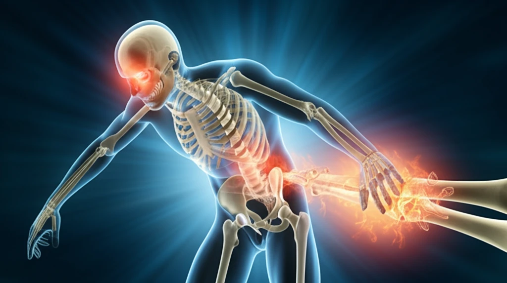
Beyond the Break: Understanding and Managing Heterotopic Ossification After Injuries

"A comprehensive guide to heterotopic ossification, its causes, symptoms, and the latest management strategies for athletes and active individuals."
For athletes and active individuals, pushing the limits is part of the game. However, with intense physical activity comes the risk of injuries. While many injuries heal as expected, sometimes the body responds in unexpected ways. One such response is heterotopic ossification (HO), a condition where bone tissue forms outside the normal skeleton.
This article dives into the specifics of HO, particularly as it relates to injuries sustained during sports and other physical activities. We'll explore what causes HO, how to recognize its symptoms, and what treatment options are available to help you get back in the game.
Whether you're a seasoned athlete, a weekend warrior, or simply someone who enjoys staying active, understanding HO can help you take proactive steps to protect your musculoskeletal health. Let's get started!
The Burden of Heterotopic Ossification
Heterotopic ossification (HO) represents a significant clinical challenge across multiple injury populations. A systematic review and meta-analysis focused on spinal cord injury found HO to be a notable complication warranting evidence-based prevention strategies. HO incidence has also been documented in patients with isolated limb fractures accompanied by mild traumatic brain injury, as well as among athletes—particularly NFL players—where an unusually high rate of HO was observed following platelet-rich plasma injections for core muscle injuries. Additionally, the condition has gained prominence in military medicine due to rising rates of combat-related extremity injuries.
Defining and Understanding HO
Heterotopic ossification is defined as the abnormal formation of extraskeletal bone within soft tissue, a pathological osteogenesis that involves osteoblasts and osteoclasts yet differs from normal bone formation in key features. The condition commonly follows traumatic brain injury, spinal cord injury, thermal injuries, surgical procedures, or repetitive microtrauma from contact sports. Neurogenic HO, driven by central nervous system injuries, represents a particularly high-risk subset. The most common sites for HO development include the hip, knee, elbow, and shoulder, though the exact molecular mechanisms driving the process remain an area of active investigation.
HO's Clinical Recognition
Heterotopic ossification has long been recognized as a condition in which bone tissue develops in soft tissues, most often following injury or surgical intervention, with rare genetic forms also documented. Comprehensive reviews have characterized HO as a diverse pathologic process involving the formation of extra-skeletal bone in muscle and soft tissues, establishing foundational definitions that continue to guide clinical understanding. These early conceptual frameworks laid the groundwork for ongoing research into the cellular and molecular drivers of ectopic bone formation.
What is Heterotopic Ossification (HO) and Why Does It Happen?
Heterotopic ossification (HO) is the process where bone tissue develops in areas where it shouldn't, such as in muscles or other soft tissues. While the exact cause of HO is not always clear, it often occurs following trauma, surgery, or neurological conditions. In the context of sports injuries, HO can develop after a direct blow to a muscle or joint, leading to inflammation and subsequent bone formation.
- Direct trauma to the muscle
- Extensive surgery, particularly hip or elbow replacements
- Neurological conditions such as spinal cord injuries or traumatic brain injuries
- Genetic predisposition
- Underlying inflammatory conditions
Advancing Knowledge in HO
Research into heterotopic ossification continues to evolve, with investigators exploring novel cellular pathways, refining animal models, and reassessing risk profiles across different patient populations. While no single breakthrough has yet transformed standard care, the cumulative body of work reflects a growing understanding of the biological complexity underlying ectopic bone formation and its varied clinical contexts.
Complexities in Trauma-Induced HO
Trauma-induced heterotopic ossification involves intricate molecular crosstalk that ultimately activates stem cells to initiate heterotopic endochondral ossification, a pathway that has proven difficult to predict or fully prevent. The development of animal models that recapitulate specific traumatic injuries has greatly advanced mechanistic understanding, yet translating these insights into reliably effective clinical interventions remains an ongoing challenge.
Comparing HO Across Contexts
Heterotopic ossification manifests differently depending on the underlying cause—whether neurogenic, traumatic, or genetic—and the specific body site involved. Comparative data across injury types and patient populations suggest that the severity and mechanism of the initial insult meaningfully influence both the likelihood and extent of ectopic bone formation, though direct head-to-head comparisons across all contexts remain limited.
Living Well and Staying Active with HO
While heterotopic ossification can present challenges, understanding the condition and taking proactive steps can help you manage symptoms and maintain an active lifestyle. By working closely with healthcare professionals, adhering to prescribed treatments, and making informed decisions about your activities, you can continue to pursue your fitness goals and enjoy a fulfilling life.
Pulling It Together
The evidence base for heterotopic ossification spans neurogenic, traumatic, and genetic etiologies, with prevention strategies still largely guided by mechanism-specific risk assessment. While pharmacological and radiation-based prophylaxis remain mainstays, emerging research points toward more targeted biological interventions. Expert consensus continues to emphasize early identification and individualized management as central to improving patient outcomes.
Emerging Therapies and Trends
A 2025 review of HO highlighted emerging potential therapies alongside conventional treatments, signaling a shift toward more targeted intervention strategies. Meanwhile, a 2026 study examining national trends in spinal cord injury-related HO hospitalizations assessed the broader healthcare system burden, noting that prior research has been limited by small sample sizes. Together, these works suggest that the field is moving toward larger-scale, system-level analyses to complement molecular and clinical investigations.
Systemic Barriers in HO Care
Despite growing awareness, heterotopic ossification continues to pose systemic challenges related to delayed diagnosis, variable prophylaxis protocols, and inconsistent access to specialized care. The absence of universally accepted screening guidelines and the heterogeneity of at-risk populations make standardization difficult. Addressing these gaps will likely require coordinated, multidisciplinary efforts spanning research, clinical practice, and health policy.
HO in Athletes and Active Populations
A study of NFL athletes between 2014 and 2019 provided a more comprehensive analysis of the association between platelet-rich plasma injections for core muscle injuries and subsequent heterotopic ossification, building on earlier alerts raised in 2015. Separately, case reports have documented HO developing after forcible physical activity, with experimental animal studies showing that strenuous exercise following immobilization can induce ectopic ossification containing periosteal tissue. These findings underscore the real-world impact of HO on athletes and physically active individuals, where treatment decisions carry direct consequences for performance and recovery.