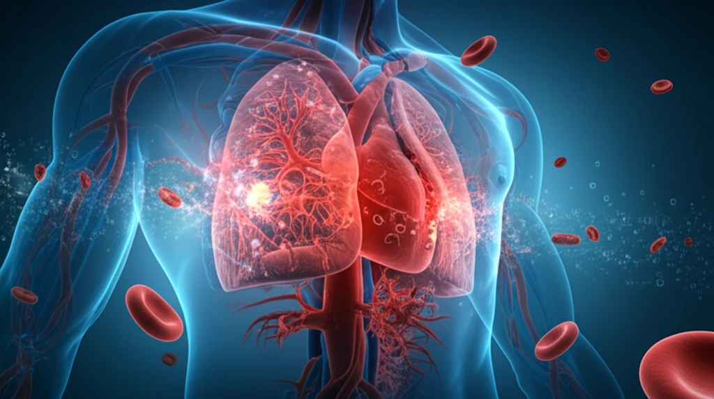
Illustration of a vein aneurysm with blood clots.

Aneurysm Alert: How a Rare Vein Condition Can Cause Life-Threatening Clots

"Unveiling the dangers of superior vena cava aneurysms, their rapid progression, and why immediate action is crucial to prevent pulmonary thromboembolism."


Aneurysms in the major veins of the chest are uncommon. Typically, they don't cause symptoms and are monitored without intervention. However, complications like rupture, blocked veins, or blood clots can occur, sometimes requiring surgery. This article discusses an unusual case of a 55-year-old man with a superior vena cava (SVC) aneurysm that rapidly worsened, leading to blood clots and a life-threatening pulmonary embolism (PTE).

This particular case underscores the importance of recognizing the potential dangers of venous aneurysms and acting swiftly when complications arise. It highlights the challenges in managing such rare conditions and the need for tailored treatment strategies.

Let’s delve into the details of this case, examining the symptoms, diagnosis, treatment decisions, and the lessons learned about managing SVC aneurysms.

The Case Unfolds: Symptoms and Initial Diagnosis

Illustration of a vein aneurysm with blood clots.

In December 2009, a 55-year-old man was admitted to the hospital with sudden chest pain, shortness of breath, and low blood pressure. He had been aware of an asymptomatic SVC aneurysm for two years, but its size had remained stable. However, upon admission, a chest X-ray revealed a significant widening in the middle of his chest, indicating rapid expansion of the aneurysm.

An echocardiogram showed that his heart’s right ventricle was enlarged and not functioning properly, suggesting pulmonary hypertension, a condition often caused by blood clots in the lungs. A computed tomographic angiography (CTA) scan confirmed the presence of a large, irregular aneurysm, measuring approximately 6.9 x 6.1 x 9.9 cm, originating from the superior vena cava and involving the left brachiocephalic vein. The aneurysm contained internal blood clots (thrombi).

  • Symptoms: Acute chest pain, dyspnea, and hypotension.
  • Previous Condition: Asymptomatic SVC aneurysm for 2 years.
  • Diagnosis: Rapidly expanding SVC aneurysm with internal thrombi, confirmed by chest radiography and CTA.
  • Echocardiogram Findings: Enlargement and hypokinesia of the right ventricle, suggesting pulmonary hypertension.
Further imaging revealed blood clots in the left pulmonary artery and its branches. Doctors considered the possibility of a hypercoagulable state (increased tendency to form blood clots), but laboratory tests ruled out any abnormalities. The initial plan was surgical removal of the aneurysm and pulmonary embolectomy (removal of blood clots from the pulmonary artery), but the patient declined surgery.

Key Takeaways for Managing SVC Aneurysms

This case highlights the critical need for vigilance in managing even asymptomatic SVC aneurysms. The rapid progression and life-threatening complications underscore the importance of considering proactive interventions and tailored treatment strategies to prevent pulmonary thromboembolism. While rare, this case serves as a reminder of the potential dangers and the need for prompt, decisive action.

About this Article -

This article was crafted using a human-AI hybrid and collaborative approach. AI assisted our team with initial drafting, research insights, identifying key questions, and image generation. Our human editors guided topic selection, defined the angle, structured the content, ensured factual accuracy and relevance, refined the tone, and conducted thorough editing to deliver helpful, high-quality information.See our About page for more information.

This article is based on research published under:

DOI-LINK: 10.3346/jkms.2011.26.5.690, Alternate LINK

Title: Unusual Cause Of Acute Right Ventricular Dysfunction: Rapid Progression Of Superior Vena Cava Aneurysm Complicated By Thrombosis And Pulmonary Thromboembolism

Subject: General Medicine

Journal: Journal of Korean Medical Science

Publisher: Korean Academy of Medical Sciences

Authors: Sang Gi Oh, Kye Hun Kim, Hyun Ju Seon, Hyun Ju Yoon, Youngkeun Ahn, Myung Ho Jeong, Jeong Gwan Cho, Jong Chun Park, Jung Chaee Kang

Published: 2011-01-01

Everything You Need To Know

1

What is a Superior Vena Cava (SVC) aneurysm, and why should I be concerned if I have one?

A Superior Vena Cava (SVC) aneurysm is a bulge or weakening in the wall of the superior vena cava, a major vein carrying blood from the upper body to the heart. While often asymptomatic and monitored without intervention, complications like rupture, vein blockage, or blood clots can arise. This case demonstrates a rapid worsening of an SVC aneurysm leading to pulmonary embolism.

2

What were the specific symptoms and diagnostic findings in the reported case of the SVC aneurysm?

The patient experienced sudden chest pain, shortness of breath (dyspnea), and low blood pressure (hypotension). An echocardiogram revealed enlargement and hypokinesia (reduced movement) of the right ventricle, suggesting pulmonary hypertension. A computed tomographic angiography (CTA) scan confirmed a large SVC aneurysm with internal blood clots (thrombi) and clots in the left pulmonary artery.

3

How does a Superior Vena Cava (SVC) aneurysm lead to a pulmonary thromboembolism (PTE), and what are the potential consequences?

A pulmonary thromboembolism (PTE) occurs when a blood clot, often originating from deep veins in the legs, travels to the lungs and blocks a pulmonary artery. In this specific case, the PTE was caused by blood clots that originated within the Superior Vena Cava aneurysm and then broke off and traveled to the pulmonary artery. This blockage can lead to reduced oxygen levels in the blood, damage to the lungs, and strain on the heart, potentially leading to life-threatening complications.

4

What is a hypercoagulable state, and why was it ruled out in this specific aneurysm case? What other factors might contribute to clot formation?

In this case, initial tests ruled out a hypercoagulable state, which is an increased tendency to form blood clots. While a hypercoagulable state can predispose someone to develop blood clots, the clots in this instance were directly linked to the SVC aneurysm itself. Further investigation may involve genetic testing or assessment for other underlying conditions that were not explicitly mentioned, but the immediate focus was on addressing the aneurysm and the existing blood clots.

5

What is the most important lesson learned from this case involving a rapidly worsening Superior Vena Cava (SVC) aneurysm, and what are the implications for managing similar conditions?

The key takeaway is the need for vigilant management of SVC aneurysms, even when asymptomatic. The rapid progression and life-threatening complications highlight the importance of considering proactive interventions and tailored treatment strategies to prevent pulmonary thromboembolism. While surgical removal of the aneurysm and pulmonary embolectomy were considered, the patient declined surgery, underscoring the challenges in managing such rare conditions. This also stresses the importance of patient education and shared decision making.

Newsletter Subscribe

Subscribe to get the latest articles and insights directly in your inbox.